HYPERSPECTRAL IMAGING
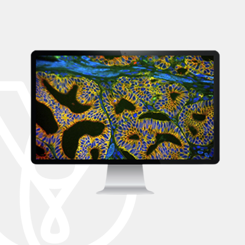
HyperSpectral imaging represents an innovative technology for a broad spectrum of applications in biomedical imaging. It combines spectroscopy, multi-dimensional imaging and computing to define the chemical composition of a biological specimen. The underlying principle is the simultaneous measurement of the detailed spectrum of every pixel in the image captured by an array sensor of the digital camera. HyperSpectral Imaging can be used to obtain Fluorescence or Brightfield spectra, such as absorption, transmission, or reflection.
HyperSpectral imaging allows for precise location of chemical constituents, providing unique and unparalleled insights into the molecular origin, formulation and phase of the observed living entity.
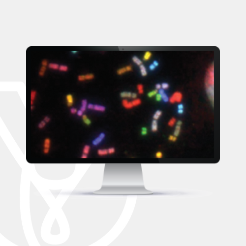
ASI’s hyperspectral solution, Rainbow, is based on Fourier Transform Spectroscopy. The system simultaneously measures the spectra at every point in the image (pixel) in the visual and low-near infrared (NIR) range. The system can be attached to any microscope.
Rainbow’s novel image analysis algorithms provide molecular and cellular image insight, beyond the visual range.
DIFFERENTIATE
Uncover chemically similar areas hidden to the eye, create color-coded maps and compare the chemical makeup of components.
SEPARATE
Separate spectral components to view them as individual layers. Detect and classify objects, based on quantitative morphological and spectral content.
QUANTIFY
Un-mix spectral components and remove background and auto-fluorescence for accurate, quantitative expression at every pixel.