PT Wadya Prima Mulia as the Authorized Distributor for ThermoFisher Scientific in Indonesia, provides Phenom ParticleX Steel Desktop Scanning Electron Microscope
Desktop SEM enabling high quality steel manufacturing through failure analysis and process improvement.
For more information regarding the product, click here
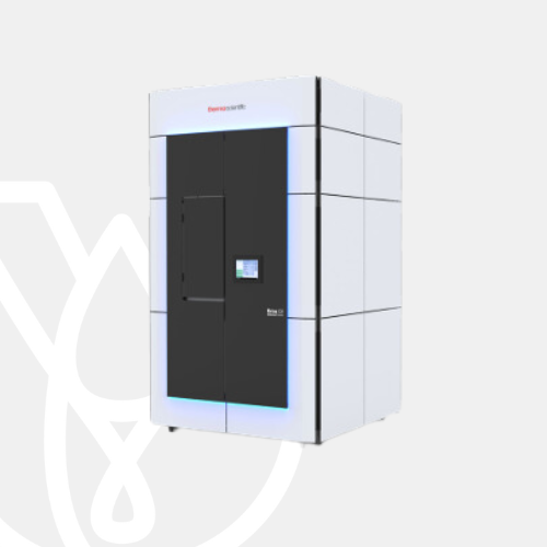
Atomic resolution cryo-EM with enhanced productivity and compact design
The award-winning Thermo Scientific Krios G4 Cryo-Transmission Electron Microscope (Cryo-TEM) enables you to unravel life at the atomic level—easier, faster, and more reliably than ever before. The most compact TEM in its class, the Krios G4 Cryo-TEM consists of a highly stable 300 kV TEM platform and the industry-leading Autoloader (a cryogenic sample manipulation robot), making it ideally suited for automated applications such as cryo-electron tomography (cryo-ET), single-particle analysis (SPA), and micro-electron diffraction (MicroED).
Solve structures at record-breaking resolutions
Pair the Thermo Scientific Krios G4 Cryo-TEM with the Thermo Scientific E-CFEG and the groundbreaking Thermo Scientific Selectris and Selectris X Imaging Filters to advance cryo-EM discovery with unprecedented resolution, speed, and ease of use. The E-CFEG is a cold field emission gun with a narrow energy spread that results in higher contrast images and enables atomic resolution. This add-on makes it possible to efficiently solve structures at resolutions of 2.0 Å or less.
Selectris Imaging Filters provide unique stability, accessibility, and performance. Paired with the Thermo Scientific Falcon 4i Direct Electron Detector, these two add-ons produce better quality images up to 10 times faster than the previous generation.
Krios G4 Cryo-TEM benefits
Truly atomic resolution performance
The optional Selectris X Imaging Filter with Falcon 4i Direct Electron Detector and E-CFEG cold field emission source improve image contrast to reach higher resolutions in shorter time.
Maximized productivity through automation
Advanced Performance Monitoring ensures the best optical starting point for automated runs and Thermo Scientific Smart EPU Software
enables automated grid screening, quality monitoring, and data management.
Workflow connectivity
Designed for easy exchange of cassettes, facilitating a smooth transfer of specimens between Autoloader-equipped TEM instruments, and equipped with Thermo Scientific Athena Software to manage experimental parameters and data.
Easier to fit into new or existing labs
Redesigned internal base frame and system enclosure reduce height below 3 m (10 ft) while enhancing system performance and preventing costly room renovations.
| Source | X-FEG (extreme high-brightness field emission gun) or low-energy-spread cold FEG (E-CFEG) |
| Accelerating voltage | 80–300 kV |
| Cryo-autoloader | Automated and contamination-free loading of cassettes (up to 12 grids) |
| Temperature management software | Includes liquid nitrogen autofill and cool down scheduling |
| Lenses | • Automatic condenser, objective and SA apertures • Three-condenser-lens system for automated, continuous, and parallel sample illumination • Symmetric constant power C-TWIN objective lens with wide-gap pole piece (11 mm) |
| Stage | • Computerized 4-axis specimen stage with ±70-degree alpha tilt • Cryo-stage with single axis holder for optimized stability and drift performance |
| Imaging | Rotation-free imaging with changing magnification |
| Advanced performance monitoring | Self-assessment of optical microscope status, combined with automated alignments, ensures ideal experimental conditions |
| Room size requirements (L × W × H) | 17’ × 22’ × 10’ |
| AFIS (aberration-free image shift) | Enhancing throughput with shorter relaxation times when moving coma-free between grid holes |
| FFI (fringe-free imaging) | Enhanced throughput with multiple image acquisitions per grid hole |
| Thermo Scientific EPU 2 Software | • Automated sample screening and data acquisition • EPU Multigrid functionality |
| Additional components | • Three 24” monitors • Hand panels to be placed within 15 meters of the column, or extend up to 300 meters from the column (optional) |
| Detectors (optional) | • Falcon 4 Direct Electron Detector • Thermo Scientific Ceta™ D Camera • Thermo Scientific Ceta 16M Camera • HAADF STEM detectors • On-axis BF/DF detectors |
| Energy filter (optional) | • Selectris Imaging Filter • Selectris X Imaging Filter • Gatan BioContinuum Energy Filter |
| Other options | • Cs Image Corrector • Thermo Scientific Phase Plate Solution |
Atomic resolution single particle analysis
Using a Krios G4 Cryo-TEM equipped with a Thermo Scientific Selectris X Imaging Filter, structural biologists achieved record-breaking resolution results, featured on the cover of Nature and other recent articles from Science and Nature. Scientists from the Medical Research Council Laboratory of Molecular Biology in Cambridge, UK, obtained a 1.2 ångström resolution structure of the iron-storing protein apoferritin. They also achieved a 1.7 ångström resolution of membrane protein (GABA receptor), with an even sharper resolution in key parts of the protein. This human membrane protein has target sites for general anesthetics, benzodiazepines, barbiturates, and neuroactive steroids, making it very relevant to structure-based drug discovery. In addition to the resolution leap, the Selectris technology can also significantly impact productivity. By having a better filter and camera, you can achieve a specific resolution with much less data.
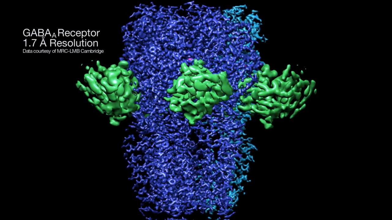
Human GABA-A receptor resolved at 1.7 Å
GABA-A is a 200 kDa human membrane protein with primary target sites for a range of clinically-relevant drugs. With the Selectris Imaging Filter, never-before-seen details of the binding pocket were resolved at 1.7 Å.

Mouse apoferritin resolved at 1.2 Å
Apoferritin is an iron-storing protein used to characterize cryo-EM performance. With the Selectris Imaging Filter, the position of individual hydrogen atoms, both in the protein and in surrounding water molecules, was resolved at 1.2 Å.

Human respiratory syncytial virus F variant
Structure of Human respiratory syncytial virus (RSV) F variant (construct pXCS847A) as described by Che et al. (PDB ID: 7UJA). The very first vaccine by GlaxoSmithKline (GSK) was approved by the Food and Drug Administration in 2023.

Janus Kinase (JAK) dimer
Structure of active Janus Kinase (JAK) dimer complexed with cytokine receptor intracellular domain as described by Glassman et al. (PDB ID: 7T6F). JAK is an essential component of cytokine signaling. Mutations to this kinase have led to immunodeficiency and myeloproliferative disorders.

SARS-CoV-2 Spike protein
Structural biology with cryo-EM has contributed to our understanding of SARS-CoV-2 since the beginning of the COVID-19 pandemic, providing critical information on the viral spike protein. Structure as described by Zhang et al. (PDB ID: 7KRQ).

TMEM106B filaments
Alzheimer’s disease associated TMEM106B filaments as described by Chang et al. (PDB ID: 7QVC). They showed that a previously unsolved amyloid fibril is composed of TMEM106B, a transmembrane lysosomal/endosomal protein.
For other products from ThermoFisher, click here.