Olympus Confocal Laser Scanning Microscope FV3000
Super Resolution Software Module
The FLUOVIEW FV3000 series of confocal laser scanning microscopes meets some of the most difficult challenges in modern science. Featuring the high sensitivity and speed required for live cell imaging as well as deep tissue observation, the FV3000 confocal microscope enables a wide range of imaging modalities, including macro-to-micro imaging, super resolution microscopy, and quantitative data analysis.
For more information about this product click here.
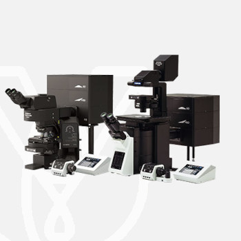
Olympus FV3000 has several configurations that can be selected according to your application.
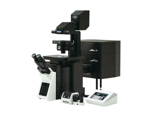
Inverted Microscope
- Suitable for observing cells cultured in a vessel
- TruFocus unit enables time-lapse observations with consistent focus
- Add a stage-top or full enclosure incubator to maintain the environmental conditions of cultured cells
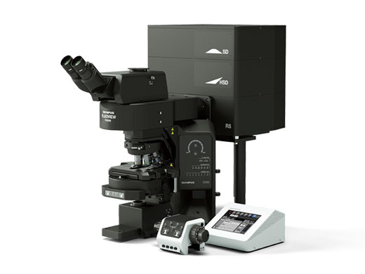
Upright microscope (configured for slide imaging)
- Optimized for fixed tissue and glass slide specimens
- Motorized 7-position nosepiece and condenser enable automated transitions from low to high magnification for macro-to-micro applications
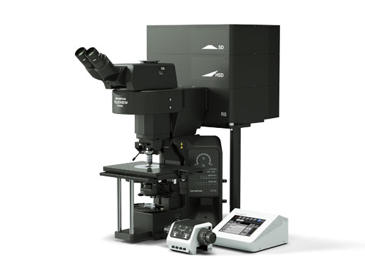
Upright microscope (configured for electrophysiology)
- Ample space around the objectives enables easy installation of patch-clamp devices
- Add extra space by lowering the stage position for experiments that require large sample handling
- Swing and slider nosepieces are available so objectives can be easily changed without interfering with the patch-clamp setup
TruSpectral High-Sensitivity Multichannel Imaging
Using proprietary spectral detection technology, the FV3000 confocal microscope’s TruSpectral detectors combine high sensitivity with spectral flexibility to detect even the dimmest fluorophores.
- Up to 3X more light transmission vs. traditional spectral detection technology
- Independently adjustable channels to optimize signal detection for each individual fluorophore
- Lambda scanning mode enables accurate spectral unmixing of complex overlapping fluorescent signals
- Variable barrier filter mode provides simultaneous four-channel image acquisition, with up to sixteen channels in virtual channel mode
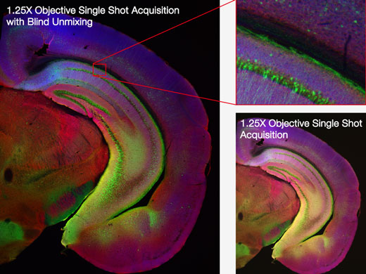
Macro-to-Micro Imaging and Super Resolution Microscopy
The FV3000 microscope’s macro-to-micro workflow provides a roadmap for data acquisition, enabling you to see data in context and easily locate regions of interest for higher resolution imaging.
- Use a low-magnification 1.25X or 2X objective to quickly capture a large field of view (FOV) map of whole specimens
- Identify regions of interest on the Overlay Map, then switch to a higher magnification objective for high-resolution confocal imaging down to 120 nm with Olympus Super Resolution technology (FV-OSR)
- Finalize your acquisition and get publication-ready microscope images with TruSight image processing
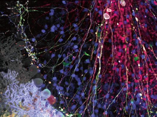
Mouse brain hemisection embedded for expansion microscopy (pre-expansion), labeled with secondary antibodies against GFP (Alexa Fluor 488, green), SV2 (Alexa Fluor 565, red), Homer (Alexa Fluor 647, blue).
Sample courtesy of Dr. Ed Boyden and Dr. Fei Chen, MIT.
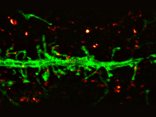
Dendrite (anti-GFP Alexa Fluor 488, green) and synaptic marker (SV2, Alexa Fluor 565, red). Olympus Super Resolution image processed with cellSens advanced constrained iterative deconvolution. Average full width half maximum measurements ~135 nm. Image acquired with 100X 1.35 NA silicone objective.
Sample courtesy of Dr. Ed Boyden and Dr. Fei Chen, MIT.
Hybrid Scanning for High-Speed Imaging and Increased Productivity
The FV3000 Hybrid Scanner provides two scanners in one for enhanced confocal imaging capabilities.
- The FV3000RS hybrid scan unit uses a galvanometer scanner for precision scanning as well as a resonant scanner, ideal for high-speed imaging of live physiological events
- Capture video-rate images with a large FOV using the resonant scanner, featuring speeds from 30 frames per second (fps) at FN 18 all the way up to 438 fps using clip scanning
- Use the resonant scanner to observe fast phenomena, such as a beating heart, blood flow, or calcium ion (Ca2+) dynamics inside cells
- Switch between the galvanometer scanner and resonant scanner with the click of a button
Accurate Time-Lapse Imaging
Time-lapse experiments require consistent focus and low phototoxicity to the sample.
- Olympus’ TruFocus unit helps maintain focus during live cell imaging despite changes in temperature or added reagents
- The FV3000 microscope’s high-sensitivity detector requires significantly less laser power while the resonant scanner reduces laser illumination time, lowering phototoxicity for more physiologically accurate confocal imaging data
Deep Tissue Observation with Silicone Objectives
The refractive index of silicone oil is close to that of living tissue, enabling high-resolution observation deep inside tissue with minimal spherical aberration.
- Refractive index match delivers an ideal focal volume, resulting in perfect volume reconstruction and enabling high-resolution confocal imaging of large living organisms
- Long working distances enable detailed microscopic imaging at depth
- See data unfold in real-time and easily observe structures with 3D reconstruction software
| FV3000 | FV3000RS | ||
| Main Laser Combiner | Violet/Visible Light Laser | 405 nm: 50 mW, 488 nm: 20 mW, 561 nm: 20 mW, 640 nm: 40 mW One optional laser port for the sub laser combiner or optional laser unit | |
| Optional Laser | Sub Laser Combiner | Maximum 3 laser units as follows: 445 nm: 75 mW, 514 nm: 40 mW, 594 nm: 20 mW, fiber connected to main laser combiner | |
| Single Laser Unit | 445 nm: 75 mW, 514 nm: 40 mW, or 594 nm: 20 mW, directly connected to main laser combiner | ||
| Laser Light Control | Main laser combiner with implemented AOTF system, ultra-fast intensity modulation with individual laser lines, additional shutter control continuously variable (0.1%–100%, 0.1% increments) 10% or 100% maximum laser power using an ND filter | ||
| Scanner | Scanning Method | 2 silver-coated galvanometer scanning mirrors | 2 silver-coated galvanometer scanning mirrors 1 silver-coated resonant and 1 silver-coated galvanometer scanning mirrors |
| Galvanometer Scanner (Normal Imaging) | Scanning Resolution: 64 × 64 to 4096 × 4096 pixels Scanning Speed (One Way): 512 × 512 with 1.1 s–264 s. pixel time: 2 µs–1000 µs Scanning Speed (Round Trip): 512 × 512 with 63 ms–250 ms, 256 × 256 with 16 ms–125 ms Optical Zoom: 1X–50X in 0.01X increments Scan Rotation: Free rotation (360 degree) in steps of 0.1 degree Scanning Mode: PT, XT, XZ, XY, XZT, XYT, XYZ, XYλ, XYZT, XYλT, XYλZ, XYλZT ROI scanning, rectangle clip, ellipse, polygon, free area, line, free line and point, tornado mode only for stimulation | ||
| Resonant Scanner (High-Speed Imaging) | – | Scanning Resolution: 512 × 32 to 512 × 512 pixels Scanning Speed: 30 fps at 512 × 512, 438 fps at 512 x 32 Optical Zoom: 1X–8X in 0.01X increments Scanning Mode: XT, XZ, XY, XZT, XYT, XYZ, XYλ, XYZT, XYλT, XYZ, XYλZT ROI scanning, rectangle clip, line | |
| Pinhole | Single motorized pinhole, pinhole diameter ø50–800 µm (1 µm steps) | ||
| Field Number (FN) | 18 | ||
| Dichromatic Mirror Turret | 8 positions (high-performance DMs and 10/90 mirror) | ||
| Optional Unit for Scanner | Laser power monitor, optional laser port | ||
| High-Sensitivity Spectral Detector | Detector Module | Cooled GaAsP photomultiplier, 2 channels | |
| Spectral Method | Motorized volume phase holographic transmission diffraction grating, motorized adjustable slit, selectable wavelength bandwidth: 1–100 nm, wavelength resolution: 2 nm | ||
| Dichromatic Mirror Turret | 8 positions (high-performance DMs and mirror) | ||
| System Control | Control Unit | OS: Windows® 7 Professional 64-bit (English version), Windows 10 Professional 64-bit ; built-in dedicated I/F board and hardware sequencer for precise imaging timing | |
| Display | 30- or 32-inch monitor (WQUXGA 2560 × 1600) | ||
| Fluorescence Illumination Unit | External fluorescence light source, fiber adaptor to the optical port of the scan unit, motorized switching between the LSM light path and fluorescence illumination | ||
| Transmitted Light Detector Unit | Module with integrated external transmitted light photomultiplier detector and LED lamp, motorized switching | ||
Microscope
| Inverted frame | Upright frame (for imaging) | Upright frame (for electrophysiology) | |
| Microscope Frame | Motorized inverted microscope IX83 (IX83P2ZF) | Motorized fixed stage upright microscope BX63L | Motorized fixed stage upright microscope BX63L |
| Revolving Nosepiece | Motorized sextuple revolving nosepiece | Motorized septuple revolving nosepiece | Coded swing nosepiece Coded slider nosepiece |
| Condenser | Motorized long working distance condenser | Motorized universal condenser | Manual long working distance condenser |
| Focus Stroke | Built-in motorized nosepiece focus Stroke: minimum increment: 0.01 μm | ||
Software
| Basic Features | GUI designed for darkroom environment. User-arrangeable layout. Acquisition parameter reload features. Hard disk recording capability; adjust laser power and HV with Z-stack acquisition. Z-stack with alpha blending, maximum-intensity projection, iso-surface rendering. |
| 2D Image Display | Each image display: single-channel side-by-side, merge, cropping, live tiling, live tile, series (Z/T/λ), LUT: individual color setting, pseudo-color, comment: graphic and text input. |
| 3D Visualization and Observation | Interactive volume rendering: volume rendering display, projection display, animation displayed. 3D animation (maximum intensity projection method, α blending) 3D and 2D sequential operation function. |
| Image Format | OIR image format 8/16-bit gray scale/index color, 24/ 32/ 48-bit color, JPEG/ BMP/ TIFF image functions, Olympus multi-tif format. |
| Spectral Unmixing | Fluorescence spectral unmixing modes (up to 16 channels) |
| Image Analysis | Region and line measurements, intensity plot over time/Z, colocalization analysis. |
| Statistical Processing | 2D data histogram display. |
| Optional Software | Motorized-stage control Mapping and multipoint simulation Sequence manager Virtual channel acquisition Microplate navigation Remote development kit Super resolution imaging (FV-OSR) Digital camera control function Deconvolution FRET and FRAP analysis Automatic object measurement and classification Object tracking |
For other products from Evident-Olympus, click here.