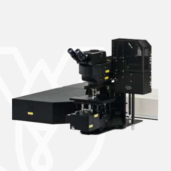
Olympus Laser Scanning Microscope FVMPE-RS
Designed for deep imaging in biological specimens, the FVMPE-RS multiphoton microscope helps reveal how cells function and interact within living tissue.
For more information about this product click here.
High-Sensitivity, High-Resolution Deep Multiphoton Imaging
The FVMPE-RS multiphoton microscope employs advanced technology and optical design to enhance sensitivity and resolution during deep imaging.
- Broad 400 nm to 1600 nm spectral transmission window efficiently delivers near-infrared excitation without compromising short wavelength detection
- Large-area detection path collects more emission signal, especially large-angle scattered photons
- TruResolution objectives offer automated spherical aberration compensation to increase brightness and resolution, revealing fine details at every plane within a deep image stack
High-Speed Imaging for Fast, Dynamic Cellular Processes
High-speed resonant scanning and high-resolution linear scanning are standard.
- Full frame imaging at 30 frames-per second (fps) reduces motion artifacts during in vivo and live cell imaging
- 438 fps maximum imaging speed enables you to capture rapid, dynamic phenomena, such cell transport during blood flow and calcium signaling events in neurons and other cells
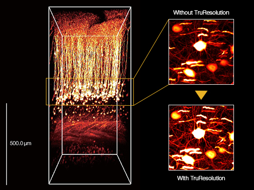
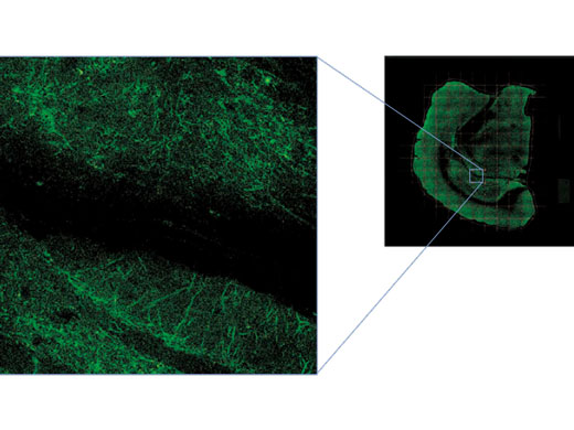
3D reconstructed image of an in vivo mouse brain (Thy1-YFP-H mouse, sensory cortex) acquired using a TruResolution objective with an auto adjustment function (left) and maximum projection images acquired at an approximately 600 μm depth. Images acquired without (top right) and with (bottom right) the auto adjustment function.
Images were acquired at the RIKEN BSI-Olympus Collaboration Center, courtesy of Dr. Hiromu Monai, Dr.Hajime Hirase, and Dr. Atsushi Miyawaki.
Multiwavelength Excitation for Broader Spectral Coverage
The FVMPE-RS imaging platform supports a dual wavelength infrared pulsed laser or two independent tunable infrared lasers for multichannel, multiphoton excitation imaging.
- Optimally excite different fluorophores without having to repeatedly tune the laser
- Independent power control of each laser line makes it easier to simultaneously capture fluorophores with differing efficiencies
- Laser wavelengths beyond 1000 nm enables access to the growing library of red-shifted fluorescent dyes and proteins and practical third-harmonic generation imaging
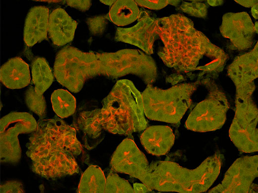
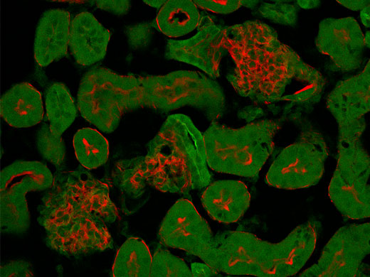
Third harmonic generation imaging of porcine adipose tissue. Unlabeled porcine adipose tissue was irradiated with femtosecond laser at 1250 nm, second harmonic is detected from collagen fibers at 625 nm and third harmonic from lipid interfaces at 416 nm.
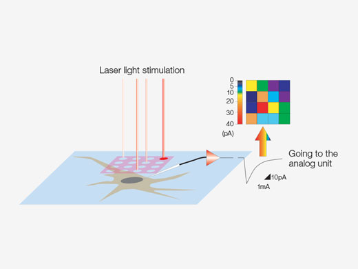
Create 3D Stimulation Reaction Maps
Specialized mapping scans utilize a pseudorandom sequence to deliver more precise spatial reaction maps when measuring electrophysiological response to optical stimulation. Direct electrical patch clamp recordings and fluorescence signals from calcium or voltage indicators can be overlaid on a high-resolution image. Intensity thresholds can be used to define a high-speed multipoint scan of the most active regions. This can be extended to 3D reaction maps with an optional piezo Z drive.
Combine a Wide Field of View with High Resolution
See your entire specimen at high resolution and in wider context with the multiarea time lapse (MATL) function:
- Automatically capture and stitch multiple adjacent images over a very wide field of view using a motorized stage
- Program time lapse acquisition over the entire composite field, or even non adjacent areas
- Easily locate the position of specific cells in the larger image with the mapping feature
Expanded Analysis Functions
The FVMPE-RS imaging platform’s software is integrated with Olympus cellSens image analysis software, expanding the system’s analytical capabilities.
Optional features include:
- 3D deconvolution for Z-stack images
- Area estimation for each particle in an image
- An image processing filter
- Colocalization analysis
Separate Overlapping Channels with Spectral Deconvolution
Closely overlapping fluorescence spectra can complicate biological studies that look at multiple labels simultaneously. Now, overlapping spectral channels can be separated using spectral deconvolution based on a blind unmixing algorithm or previously saved multichannel profiles. Crosstalk between the channels can even be eliminated during image acquisition through live processing.
Normal Mode
Live Unmixing Mode
FLUOVIEW FVMPE-RS
| One Laser System | Dual Lines System | Twin Lasers System | ||
| Unit | Qualified IR Pulsed Lasers with Negative Chirp for Multiphoton Excitation | Spectra-Physics products: MAITAI HPDS-OL: 690 nm–1040 nm MAITAI eHPDS-OL: 690 nm–1040 nm INSIGHT X3-OL: 690 nm–1300 nm INSIGHT X3 DUAL/DUALC-OL: 680 nm–1300 nm + 1045 nm Coherent products: Chameleon Vision I Olympus: 680 nm–1080 nm Chameleon Vision II Olympus: 680 nm–1080 nm Chameleon Vision I Olympus: 690 nm–1050 nm | ||
| Main IR Pulsed Laser | MAITAI HPDS-OL MAITAI eHPDS-OL INSIGHT X3-OL Chameleon Vision I Olympus Chameleon Vision II Olympus Chameleon Vision S Olympus | INSIGHT X3 DUAL-OL INSIGHT X3 DUALC-OL | MAITAI HPDS-OL MAITAI eHPDS-OL INSIGHT X3-OL Chameleon Vision I Olympus Chameleon Vision II Olympus Chameleon Vision S Olympus | |
| Additional IR Line Laser: Use as second imaging line/laser or for simultaneous stimulation (optional SIM scanner) | – | 1045 nm fixed line from INSIGHT X3 DUAL /DUALC-OL | MAITAI HPDS-OL MAITAI eHPDS-OL Chameleon Vision I Olympus Chameleon Vision II Olympus Chameleon Vision S Olympus | |
| Automatic Introduction Optic | Introduction optic with AOM attenuation (0%–100%, 0.1% increments) Including fully automated beam expander, XY shifter and two-axis angle alignment. (4-axis quadralign autoalignment optics) Direct coupling to laser port of scanning unit. | Introduction optic with 2 sets of AOM attenuation (0%–100%, 0.1% increments) Including 2 sets of fully automated beam expanders, XY shifter and two-axis angle alignment. (4-axis quadralign autoalignment optics) Direct coupling to laser port of scanning unit. | ||
| IR Laser Combining Optic | – | Motorized light path switcher with DM900, DM1000R, DM1100 to combine two IR wavelength for imaging | ||
| Optional Visible Light Laser for Stimulation | 405 nm/50 mW, 458 nm/20 mW, 588 nm/20 mW laser source with AOTF attenuation. 0%–100%, 0.1% increments, < 2 μs rising time | |||
| Scanning Unit | Scanning Method | Light deflection via 2 silver-coated galvanometer scanning mirrors, or silver-coated resonant scanning mirror | ||
| Scanning Speed | Galvanometer scanner (normal imaging): 512 × 512 with 1.1 s–264 s, pixel time: 2 μs–1000 μs Resonant scanner (high-speed imaging): 30 fps at 512 × 512, 438 fps at 512 × 32 | |||
| Scanning Mode | XY, XYZ, XYT, XYZT, free line, XZ, XT, XZT, PointT | |||
| Galvanometer Scanner (Normal Imaging) | Galvanometer ROI scanning: rectangle clip, ellipse, polygon, free area, line, free line, and point Zoom: 1.0x–50.0x with 0.01x increments, supports 0°–360° rotation and pan Scanning Field Number: 18 Image Size: 64 × 64–4096 × 4096 | |||
| Resonant Scanner (High-Speed Imaging) | Resonant ROI scanning: rectangle clip, line Zoom: 1.0x–8.0x with 0.01x increments Scanning field number:18 Image size: 512 × 512 | |||
| Optical Coating | IR support optic with 1600 coating | |||
| Non-descanned MPE Imaging Detectors | Reflected detection: 2 or 4 channel configuration: 2 PMT configuration, 4 PMT configuration or 2 PMT + 2 cooled GaAsP-PMTs Transmitted detection: 2 PMT unit with high NA condenser | |||
| Transmitted-Light Detector | Module with integrated external transmitted light photomultiplier detector and 100 W halogen lamp, motorized switching, fiber adaptation to microscope frame | |||
| Z-Drive | Integrated motorized focus module of the microscope, minimum increment 0.01 μm Optional: highly rigid piezo nosepiece*1 | |||
| Optional Simultaneous Stimulation Scanner | Highly synchronized simultaneous stimulation scanner, including a set of galvanometer scanner, VIS and IR laser port. ROI scanning: rectangle clip, ellipse, polygon, tornado, free area, line, free line, and point. | |||
| Optional Analog and Digital in/out Box | 4-channel analog signal input, 6-channel digital TTL trigger input, 5-channel digital TTL trigger output. Scanner timing output | |||
| Operation Environment | Room temperature: 20°C–25°C, humidity: 75% or less at 25°C, requires continuous (24-hour) power supply | |||
| Size of Anti-Vibration Table | 1500 mm × 1650* mm *1800 mm with inverted microscope system | 1500 mm × 1650* mm *1800 mm with inverted microscope system | 1500 mm × 2000 mm | |
| Software | Basic Feature | Dark room matching GUI design. User arrangeable layout. Acquisition parameter reload features. Hard disk recording capability, Adjust laser power and HV with Z-stack acquisition. Z-stack with alpha blending, Maximum intensity projection, Iso-surface rendering | ||
| IR Laser Control | Fully integrated IR laser wavelength control and Deep Focus mode | |||
| Optional Motorized Stage software | XY motorized stage control. Map image acquisition for easy target locating. Tiling acquisition and software image stitching. Define multiple area for time lapse imaging. | |||
| Optional Mapping and Multiple Point Stimulation Software | Multiple point stimulation and data acquisition software. Mapping multiple point stimulation to generate reaction map. Filtering feature to select points. Multiple point stimulation. Single or repeat stimulation. Each point independent stimulation wavelength selection. | |||
| Optional Sequence Manager | Advanced programmable software to define multiple imaging/stimulation tasks and execute by hardware sequencer. Minimum gap 100 ms delay between tasks. | |||
| Optional Auto Compensation Software | Automatic spherical aberration compensation software. Control of objective lens with auto spherical aberration compensation function. Auto adjustment of motorized correction collar to find the best position at certain observation depth. Auto adjustment of correction collar along with Z movement. | |||
*1 Not available in some areas.
TruResolution Objectives
| FV30-AC10SV | FV30-AC25W | |
| Magnifications | 10 | 25 |
| NA | 0.6 | 1.05 |
| W.D. | 8 mm | 2 mm |
| Cover Glass Thickness | 0 mm–0.23 mm | 0 mm–0.23 mm |
| Immersion Liquid | SCALEVIEW-A2 (water, silicone oil, and normal oil available) | Water |
| Special Features | Auto compensation, optimized for multiphoton imaging | Auto compensation, optimized for multiphoton imaging |
| Dimensions (W × D × H) | 56 mm × 106.5 mm × 95 mm | 56 mm × 106.5 mm × 101 mm |
| Weight | Approx. 1kg | Approx. 1kg |
For other products from Evident-Olympus, click here.