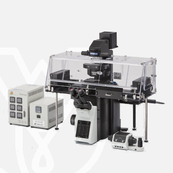
Olympus Inverted Microscope IXplore Live
PT Wadya Prima Mulia as the Exclusive Distributor for Evident-Olympus in Indonesia, provides Inverted Microscope IXplore Live and others.
Precise Live Cell Imaging
Designed for precise live cell imaging, the IXplore Live microscope system helps reduce photobleaching and enhance cell viability for physiological experiments.
- Utilize the Olympus real-time controller for physiologically relevant data with minimal cell disturbance
- Maintain cell viability while imaging with various environmental control options
- Maintain focus accurately and reliably in time-lapse experiments with TruFocus
- Discover the real structure of your cells with Olympus silicone oil immersion optics
For more information about this product click here.
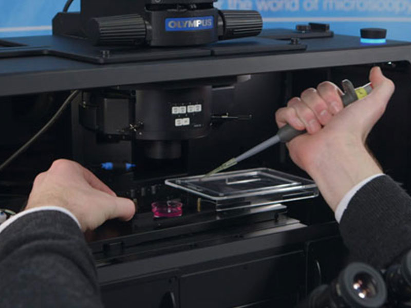
Meeting the Needs of Live Samples
Live cells require careful maintenance of their environment to grow and thrive, and Olympus offers a variety of microscope-based incubation systems to meet evolving research needs. Box-type incubation systems* enable time-lapse observations over a period of several days by enclosing a portion of the microscope within the incubator. Shorter time-lapse experiments can be completed with stage-top microscope CO2 incubation systems* that can be fitted to the stage and easily removed when not in use.
Both systems are precisely controlled to maintain a constant environment surrounding the dish or well plates, controlling temperature, humidity, and CO2 concentration. This maintains cell activity to significantly improve the reliability of time-lapse observation and provide better data.
*Third-party products.
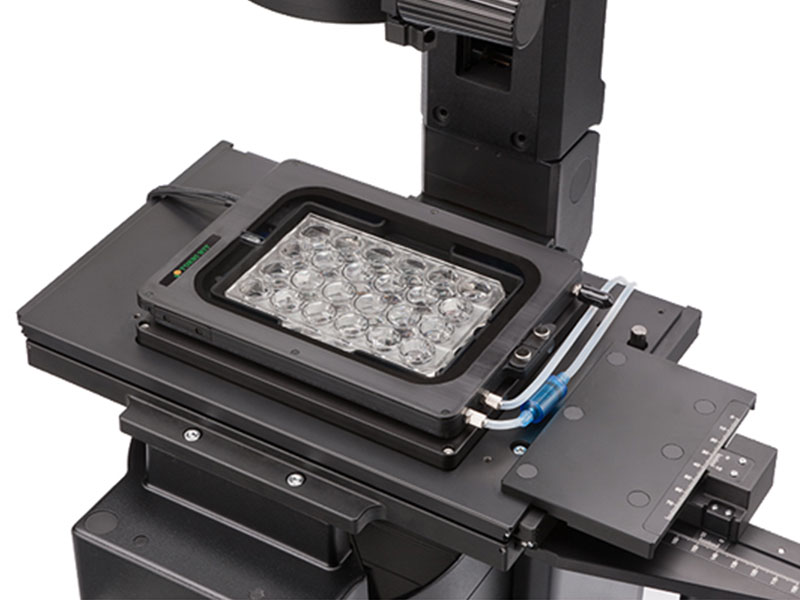
Hardware Stability
The frame architecture and focus drive design of the IXplore system offer enhanced rigidity that reduces the impact of vibration and temperature on the microscope. This helps maintain desired positions along the X, Y, and Z axes to facilitate reliable time-lapse and multipoint imaging. When using the IXplore Live system combined with the Olympus ultrasonic stage (IX3-SSU) and TruFocus system, you can capture high-precision, multipoint time-lapse images that are aligned and in focus.
Live Cell Imaging
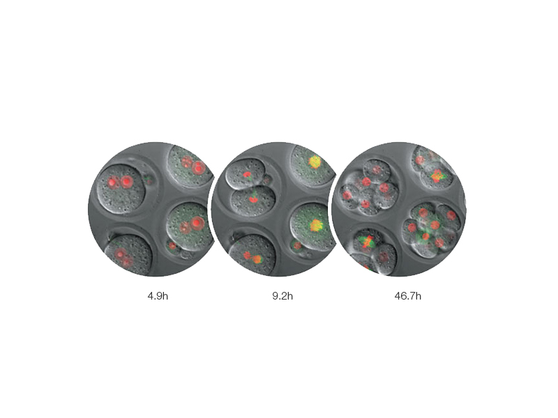
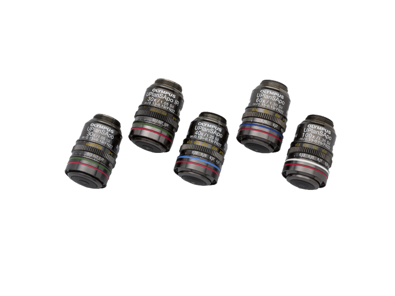
Olympus silicone oil immersion objectives provide clearer images of living specimens during time-lapse experiments. The refractive index of silicone oil (ne≈1.40) is close to that of living tissue (ne≈1.38), so these objectives help reduce the spherical aberration caused by refractive index mismatch, enabling high-resolution observation deep inside living tissue. Silicone oil does not dry out or harden, so there is no need to add more oil, making it ideal for extended time-lapse observations.
Monitor Cell Migration and Growth
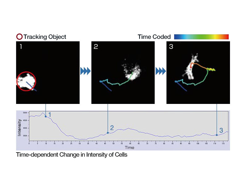
Analyze the movement and division of live cells in time-lapse or z-stack image sets with cellSens Object Tracking and Count and Measure solutions. Use the Confluency Checker tools to measure confluency on phase contrast images in addition to fluorescence.

Rapid Deconvolution
Olympus cellSens Dimension software includes live 2D deblurring for preview and acquisition to enable better focusing on thick specimens. More advanced TruSight deconvolution is available to reassign out-of-focus light through the CI deconvolution solution. TruSight uses a constrained iterative deconvolution algorithm to produce improved resolution, contrast, and dynamic range with high speed through GPU processing.
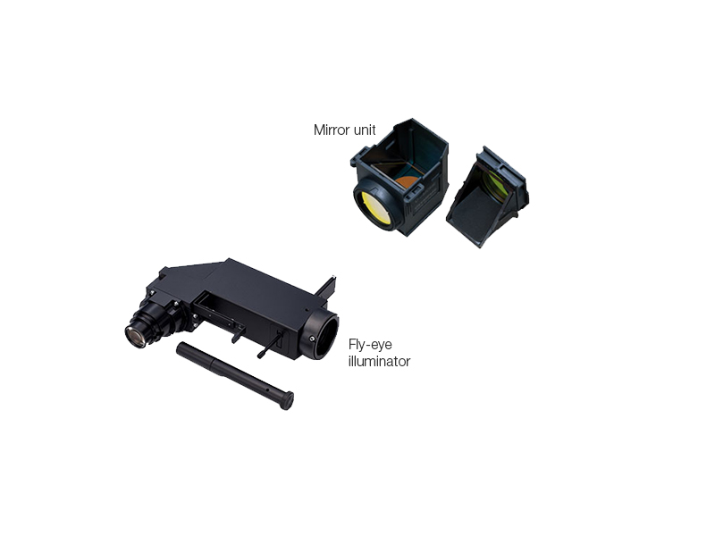
Large Field of View
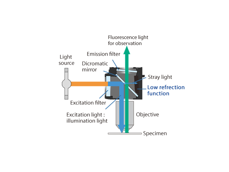
The large field of view Olympus optics, including mirror units and fly-eye lens systems, provide uniformly illuminated fluorescence images and enable the use of sCMOS cameras with large sensors.
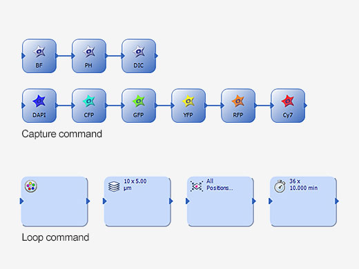
Ease of Use
The Graphical Experimental Manager (GEM) of cellSens Dimension software offers fully automated multidimensional observation (X, Y, Z, T, wavelength, and positions) and eases experiment setup.
References
S. Wakayama, et al. Chemical labelling for visualizing native AMPA receptors in live neurons. Nature Communications (April 7, 2017).
S. N. Cullati, et al. A bifurcated signaling cascade of NIMA-related kinases controls distinct kinesins in anaphase. The Journal of Cell Biology (June 19, 2017).
L. Gheghiani, et al. PLK1 activation in late G2 sets up commitment to mitosis. Cell Reports (June 6, 2017).
D. Nakane and T. Nishizaka, et al. Asymmetric distribution of type IV pili triggered by directional light in unicellular cyanobacteria. PNAS (June 5, 2017).
T. A. Redchuk, et al. Near-infrared optogenetic pair for protein regulation and spectral multiplexing. Nature Chemical Biology (March 27, 2017).
S. Barzilai, et al. Leukocytes breach endothelial barriers by insertion of nuclear lobes and disassembly of endothelial actin filaments. Cell Reports (January 17, 2017).
J. Humphries, et al. Species-independent attraction to biofilms through electrical signaling. Cell (January 12, 2017).
A. Prindle, et al. Ion channels enable electrical communication in bacterial communities. Nature (October 21, 2015).
K. G. Harris, et al. RIP3 regulates autophagy and promotes coxsackievirus B3 infection of intestinal epithelial cells. Cell Host & Microbe (August 13, 2015).
| Microscope Frame |
IX83P2ZF |
||
|---|---|---|---|
| Observation Method | Fluorescence (Blue/Green Excitation) | ||
| Fluorescence (Ultraviolet Excitation) | |||
| Differential Interference Contrast (DIC) | |||
| Phase Contrast | |||
| Brightfield | |||
| Revolving Nosepiece | Motorized (6 position) | ||
| Focus | Motorized | ||
| Z Drift Compensator | |||
| Observation Tubes | Widefield (FN 22) | Tilting Binocular | |
| Illuminator | Transmitted Köhler Illuminator | LED Lamp | |
| 100 W Halogen Lamp | |||
| Fluorescence Illuminator | 100 W Mercury Lamp | ||
| Light Guide Illumination | |||
| Fluorescence Mirror Turret | Motorized (8 position) | ||
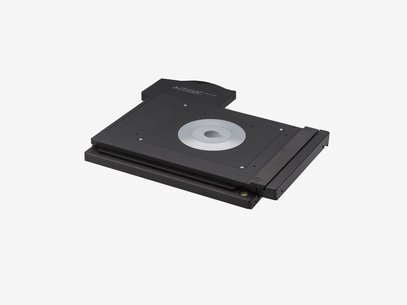
| Stage | Motorized | IX3-SSU Ultrasonic Stage for IX3 | X: 114 mm, Y: 75 mm |
| Mechanical | IX3-SVR Mechanical Stage with Right Handle | X: 114 mm, Y: 75 mm | |
| IX3-SVL Mechanical Stage with Left Short Handle | X: 114 mm, Y: 75 mm | ||
| Condenser | Motorized | Universal Condenser | W.D. 27 mm, NA 0.55, motorized aperture and polarizer |
| Manual | Universal Condenser | NA 0.55/ W.D. 27 mm | |
| Ultra-Long Working Distance Condenser | NA 0.3/ W.D. 73.3 mm | ||
| Confocal Scanner |
– |
||
| Super Resolution Processing |
– |
||
| Accessories |
Remote correction collar controller (IX3-RCC) |
||
| Dimensions (W × D × H) | 323 (W) x 475 (D) x 706 (H) mm (IX83 microscope frame) | ||
| Weight | Approx. 47kg (IX83P2ZF) | ||
For other products from Evident-Olympus, click here.