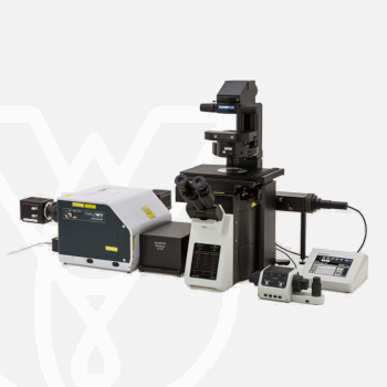
Olympus Inverted Microscope IXplore SpinSR
PT Wadya Prima Mulia as the Exclusive Distributor for Evident-Olympus in Indonesia, provides Inverted Microscope IXplore SpinSR and others.
Confocal Super Resolution for All Live Cell Samples
Designed for fast 3D super resolution imaging and prolonged cell viability in time-lapse experiments, the IXplore SpinSR microscope system offers XY resolution down to 120 nm without the need for dedicated labeling procedures.
- Super resolution down to 120nm XY resolution
- Prolonged cell viability in confocal time-lapse imaging due to less phototoxicity and bleaching
- Switch between widefield, confocal and super-resolution with one click
- Accurate 3D reconstruction with Olympus silicone oil immersion objectives
For more information about this product click here.
High-Level Super Resolution
Resolve confocal images down to 120 nm XY resolution using the confocal technique and Olympus super resolution (OSR).
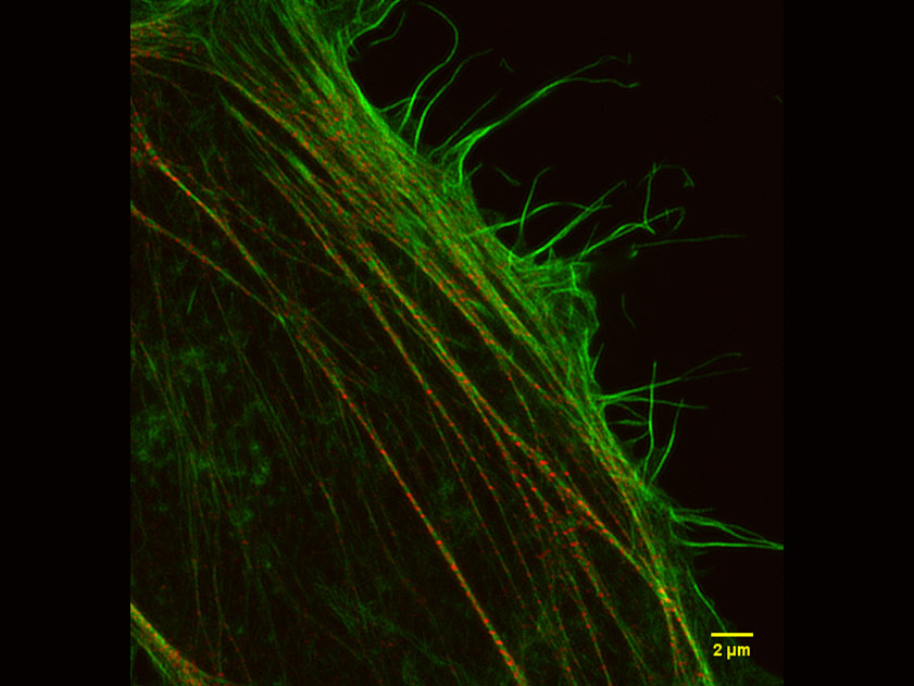
*Image: Stress fibers of Hela cell: Antibody staining with Phalloidin-Alexa488 (green) for actin, Alexa 568 (red) for myosin heavy chain.
Image data courtesy of: Keiju Kamijo,Ph.D. Division of Anatomy and Cell Biology, Faculty of Medicine, TOHOKU Medical and Pharmaceutical University
Suited for Live Specimens
OSR algorithms work in real time to eliminate delays caused by frame averaging or image reconstruction, providing instant super resolution images so you can get to your results faster.
This enables experimental design for super resolution to include live cell experiments, which are further improved through the ultrafast imaging speeds and multichannel acquisition capabilities of the spinning disk confocal.
Streamline Your Research
Easily integrate the IXplore SpinSR microscope system into existing experiment and sample protocols; you can switch from widefield, confocal, and super resolution using the same samples with just the click of a button— the microscope takes care of the rest.
Image data can be further enhanced using cellSens software’s image analysis tools. The software’s efficient workflows enable users to effectively manage their data and perform advanced analysis that helps unlock new insights.
TruSight deconvolution algorithms are designed to work seamlessly with OSR algorithms, helping prevent over processing. Together they provide sharper, clearer images than either technique alone.
Reveal Super Resolution Details Inside Your Samples
To observe intracellular structures, it is necessary to prevent out of focus fluorescence from blurring the true data in your final image. The IXplore SpinSR microscope system has incorporated a confocal optical system, enabling the use of various objectives, including silicone oil optics, therefore allowing the acquisition of sharp super resolution images with less blurring even in thick samples. Furthermore, no special fluorophores or imaging buffers are required so there is no need to change samples or preparation protocols when looking to convert to super resolution.
Reveal Your Data
Our TruSight deconvolution can be combined with super resolution to provide images that clearer and sharper than with deconvolution alone. The 3D constrained iterative deconvolution removes blur in the Z-axis for a cleaner three-dimensional image.
- High-speed image processing with the advanced TruSight deconvolution
- TruSight deconvolution is compatible with Olympus Super Resolution
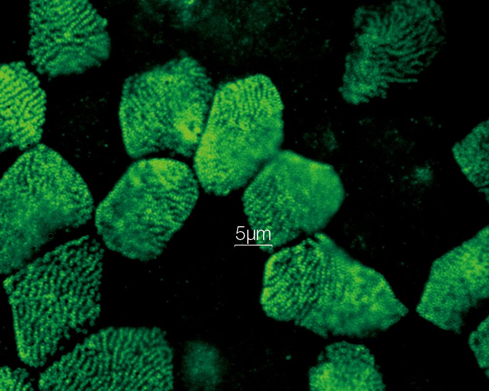
*Image: Mouse kidney tissue stained with Alexa488
Two-Color Simultaneous Imaging
The IXplore SpinSR system can use two cameras simultaneously to achieve fast, two-color localization super-resolution imaging. This is achieved using existing fluorophores and laser lines.
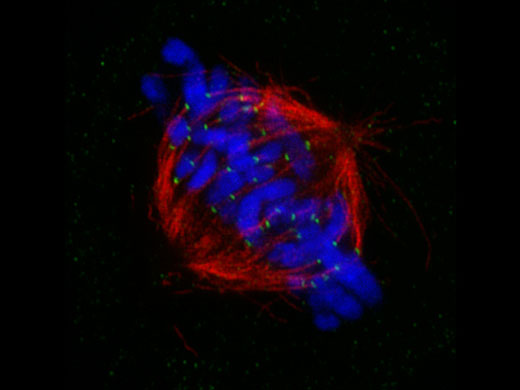
Mitotic spindle at metapahse cell
HeLa cells derived from human cervical cancer were fixed and stained for α-tublin(microtubules,red) and Hec1(kinetochores, green),respectively. DNA was stained with DAPI(chromosomes,blue). Chromosomes interact with microtubules constituting mitotic spindle via kinetochores assembled on centromere region of chromosomes.
Image courtesy of: Masanori Ikeda and Kozo Tanaka, Department of molecular oncology,Institute of Development, Aging and Cancer
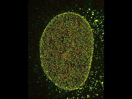
Nuclear pore complex of Hela cell
Nup153(Alexa 488: green), Nup62(Alexa 555: red)
Image courtesy of: Hidetaka Kosako, Fujii Memorial Institute of Medical Sciences, Tokushima university
References
S. Hayashi and Y. Okada, “Ultrafast superresolution fluorescence imaging with spinning disk confocal microscope optics,” Mol. Biol. Cell 26(9), 1743–1751 (2015).
S. Hayashi, “Resolution doubling using confocal microscopy via analogy with structured illumination microscopy,” Jpn. J. Appl. Phys. 55(8), 082501 (2016).
A. Nagasawa-Masuda and K. Terai, “Yap/Taz transcriptional activity is essential for vascular regression via Ctgf expression and actin polymerization,” PLoS ONE 12(4), e0174633 (2017).
H. Nakajima, et al., “Flow-Dependent Endothelial YAP Regulation Contributes to Vessel Maintenance,” Dev. Cell 40(6), 523-536.e6 (2017).
K. Tateishi, et al., “Three-dimensional Organization of Layered Apical Cytoskeletal Networks Associated with Mouse Airway Tissue Development,” Sci. Rep. 7, 43783 (2017).
E. Herawati, et al., “Multiciliated cell basal bodies align in stereotypical patterns coordinated by the apical cytoskeleton,” J. Cell Biol. 214(5) 571-586 (2016).
M.-T. Ke, et al., “Super-Resolution Mapping of Neuronal Circuitry With an Index-Optimized Clearing Agent,” Cell Rep. 14(11) 2718–2732 (2016).
| Microscope Frame |
IX83P2ZF |
||
|---|---|---|---|
| Observation Method | Super Resolution | ||
| Confocal | |||
| Fluorescence (Blue/Green Excitation) | |||
| Fluorescence (Ultraviolet Excitation) | |||
| Differential Interference Contrast (DIC) | |||
| Phase Contrast | |||
| Brightfield | |||
| Revolving Nosepiece | Motorized (6 position) | ||
| Focus | Motorized | ||
| Z Drift Compensator | |||
| Observation Tubes | Widefield (FN 22) | Tilting Binocular | |
| Illuminator | Transmitted Köhler Illuminator | LED Lamp | |
| 100 W Halogen Lamp | |||
| Fluorescence Illuminator | 100 W Mercury Lamp | ||
| Light Guide Illumination | |||
| Fluorescence Mirror Turret | Motorized (8 position) | ||
| Stage | Motorized | IX3-SSU Ultrasonic Stage for IX3 | X: 114 mm, Y: 75 mm |
| Mechanical | IX3-SVR Mechanical Stage with Right Handle | X: 114 mm, Y: 75 mm | |
| IX3-SVL Mechanical Stage with Left Short Handle | X: 114 mm, Y: 75 mm | ||
| Condenser | Motorized | Universal Condenser | W.D. 27 mm, NA 0.55, motorized aperture and polarizer |
| Manual | Universal Condenser | NA 0.55/ W.D. 27 mm | |
| Ultra-Long Working Distance Condenser | NA 0.3/ W.D. 73.3 mm | ||
| Confocal Scanner |
CSU-W1 |
||
| Super Resolution Processing |
Olympus super resolution (OSR) filter |
||
| Accessories |
Remote correction collar controller (IX3-RCC) |
||
| Dimensions (W × D × H) | 323 (W) x 475 (D) x 706 (H) mm (IX83 microscope frame) | ||
| Weight | Approx. 47kg (IX83P2ZF) | ||
For other products from Evident-Olympus, click here.