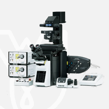
Olympus Inverted Microscope IXplore TIRF
PT Wadya Prima Mulia as the Exclusive Distributor for Evident-Olympus in Indonesia, provides Inverted Microscope IXplore TIRF and others.
Excellent Multicolor TIRF Imaging
Designed for membrane dynamics, single-molecule detection, and colocalization experiments, the IXplore TIRF microscope system offers simultaneous multicolor TIRF imaging for up to 4 colors with high stability.
- Excellent simultaneous multicolor TIRF for investigation of membrane dynamics and single molecule detection
- Exact colocalization of up to four markers thanks to individual penetration depth control
- Take advantage of Olympus’ remarkable TIRF objective with the world’s highest NA of 1.7*
- Intuitive set-up of complex experiments with Graphical Experiment Manager (GEM)
For more information about this product click here.
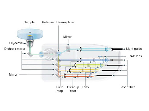
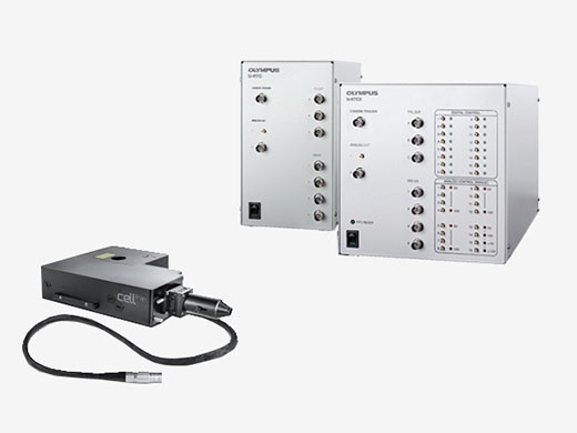
Simultaneous Multicolor TIRF
The Olympus cellTIRF system provides true simultaneous acquisition of up to four wavelengths.
- Motorized angle control for each laser provides equal evanescence (±1 nm) penetration, enabling high-contrast images of cell surface and single molecules and minimizing background noise
- cellTIRF-4Line system has integrated point FRAP optics for the first laser line, enabling FRAP and photo-switching experiments with no additional costs
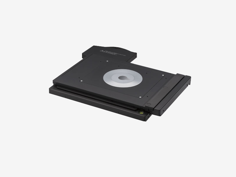
Imaging Stability
The frame architecture and focus drive design of the IXplore system offer enhanced rigidity that reduces the impact of vibration and temperature. It maintains desired positions along the X-, Y-, and Z-axes to facilitate reliable time-lapse and multipoint imaging. When combined with the Olympus ultrasonic stage (IX3-SSU) and TruFocus system, the IXplore TIRF microscope system can capture high-precision, multipoint time-lapse images that are aligned and in focus.
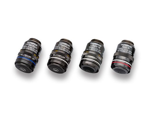
TIRF Objectives
Total internal reflection fluorescence (TIRF) is facilitated by a wide range of objectives featuring a high signal-to-noise ratio and a correction collar to adjust for cover glass thickness and temperature. Our corrected plan apochromat objectives with an NA of 1.5 help you acquire uniform high-quality images with a large field of view. Take advantage of Olympus’ remarkable TIRF objective with the world’s highest NA, 1.7*.
*As of November, 2018. According to Olympus research.
Precise and Intuitive Photomanipulation (Optional Peripherals)
Olympus’ cellFRAP photomanipulation device and real-time controller (U-RTCE) enable accurate control (200 µs dead time), diffraction-limited stimulation with a flexible region of interest, and precise reproduction of experimental conditions.
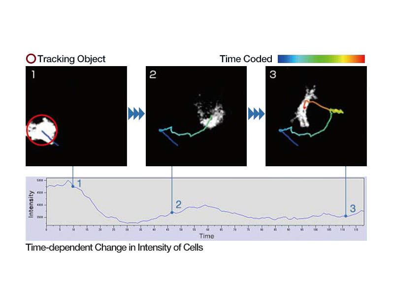
Advanced Analysis
Advanced functionality, such as object tracking, Count and Measure, Kymograph and real time 2D deconvolution to enhance object identification.
References
Y. Yang, et al. Spectraplakin induces positive feedback between fusogens and the actin cytoskeleton to promote cell-cell fusion. Developmental Cell (April 10, 2017).
A. R. van Vliet, et al. The ER stress sensor PERK coordinates ER-plasma membrane contact site formation through interaction with filamin-A and F-actin remodeling. Molecular Cell (Feburuary 23, 2017).
F. Hertel, et al. RefSOFI for mapping nanoscale organization of protein-protein interactions in living cells. Cell Reports (December 31, 2015).
C. Cauvin, et al. Rab35 GTPase triggers switch-like recruitment of the lowe syndrome lipid phosphatase OCRL on newborn endosomes. Current Biology (December 24, 2015).
W.-K. Ji, et al. Actin filaments target the oligomeric maturation of the dynamin GTPase Drp1 to mitochondrial fission sites. eLIFE (November 26, 2015).
A. Juanes-Garcia, et al. A regulatory motif in nonmuscle myosin II-B regulates its role in migratory front–back polarity. The Journal of Cell Biology (April 13, 2015).
D. Borrenberghs, et al. HIV virions as nanoscopic test tubes for probing oligomerization of the integrase enzyme. ACS Nano (March 21, 2014).
S. Yamaoka, et al. Identification and dynamics of arabidopsis adaptor protein-2 complex and its involvement in floral organ development. The Plant Cell (August 23, 2013)
| Microscope Frame |
IX83P2ZF |
||
|---|---|---|---|
| Observation Method | Total Internal Reflection Fluorescence | ||
| Fluorescence (Blue/Green Excitation) | |||
| Fluorescence (Ultraviolet Excitation) | |||
| Differential Interference Contrast (DIC) | |||
| Phase Contrast | |||
| Brightfield | |||
| Revolving Nosepiece | Motorized (6 position) | ||
| Focus | Motorized | ||
| Z Drift Compensator | |||
| Observation Tubes | Widefield (FN 22) | Tilting Binocular | |
| Illuminator | Transmitted Köhler Illuminator | LED Lamp | |
| 100 W Halogen Lamp | |||
| Fluorescence Illuminator | 100 W Mercury Lamp | ||
| Light Guide Illumination | |||
| Fluorescence Mirror Turret | Motorized (8 position) | ||
| Stage | Motorized | IX3-SSU Ultrasonic Stage for IX3 | X: 114 mm, Y: 75 mm |
| Mechanical | IX3-SVR Mechanical Stage with Right Handle | X: 114 mm, Y: 75 mm | |
| IX3-SVL Mechanical Stage with Left Short Handle | X: 114 mm, Y: 75 mm | ||
| Condenser | Motorized | Universal Condenser | W.D. 27 mm, NA 0.55, motorized aperture and polarizer |
| Manual | Universal Condenser | NA 0.55/ W.D. 27 mm | |
| Ultra-Long Working Distance Condenser | NA 0.3/ W.D. 73.3 mm | ||
| Confocal Scanner |
– |
||
| Super Resolution Processing |
– |
||
| Accessories |
Remote correction collar controller (IX3-RCC) |
||
| Dimensions (W × D × H) | 323 (W) x 475 (D) x 706 (H) mm (IX83 microscope frame) | ||
| Weight | Approx. 47kg (IX83P2ZF) | ||
For other products from Evident-Olympus, click here.