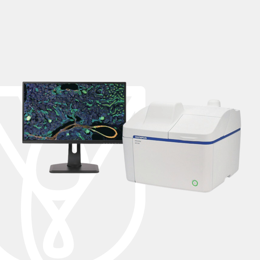
Olympus Digital Imaging System APX100
PT Wadya Prima Mulia as the Exclusive Distributor for Evident-Olympus in Indonesia, provides APX100 Digital Imaging System and other products.
The APEXVIEW APX100 all-in-one microscope makes it fast and simple to acquire expert-quality microscope images. Built with renowned Olympus optics, an intuitive user interface, a powerful AI, and a suite of smart features, the APX100 system combines the ease of use of an all-in-one-microscope with high-quality image data to fit your research needs.
Stay Focused on Your Research
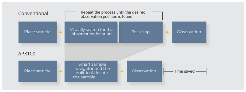
The APX100 all-in-one microscope’s smart sample navigator and fast autofocus enable you to spend less time searching for samples and more time collecting data.
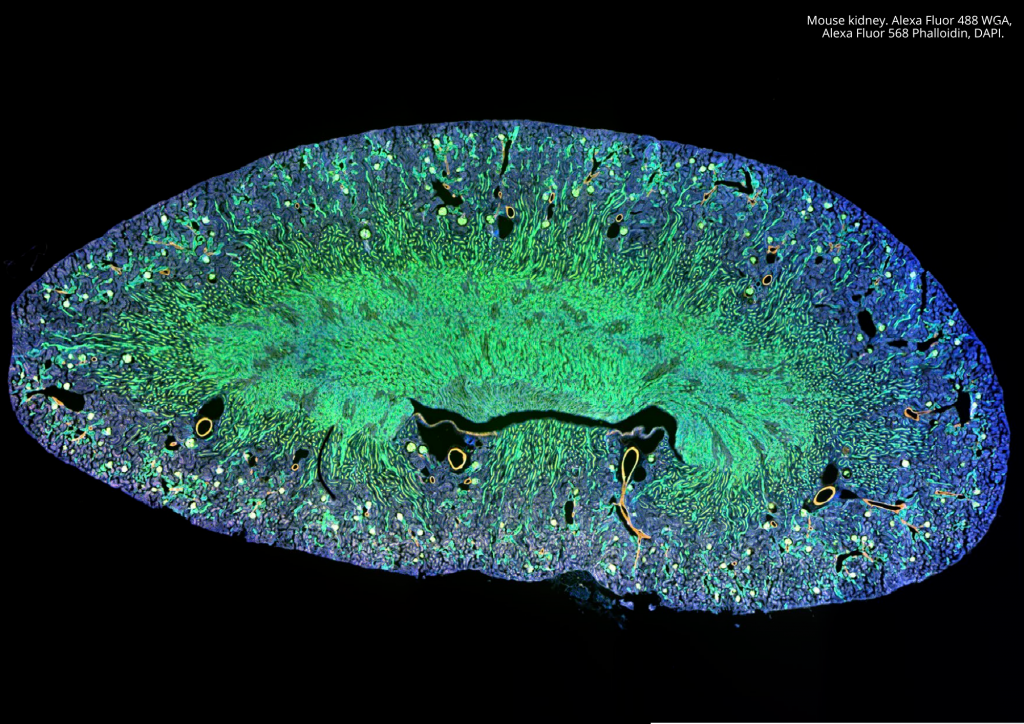
Publication-Quality Images in a Few Clicks
The APX100 microscope is built with the same quality optics and technology as our high-end systems, so you can be confident in the quality of your images.
Fast, Efficient Microscope Data Management
The system keeps your data organized and acquisition parameters saved for future reference.
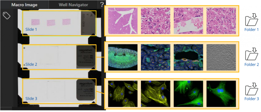
Automatic Sample Detection
Place your sample in the sample holder, and the smart sample navigator automatically acquires a macro image. The AI then locates your sample on the slide and automatically positions it above the objective, so that you can choose the observation method and immediately begin capturing images.
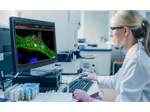
Simple Layout and Workflow Maximize Efficiency
Designed for simplicity, using the APX100 microscope is as easy as loading your sample, closing the lid, and pushing a button.
- Clear software layout and streamlined workflow
- Begin imaging with very little training
High-Speed Autofocus
With an autofocus up to twelve times faster than conventional autofocus algorithms, you can locate the imaging plane quickly.
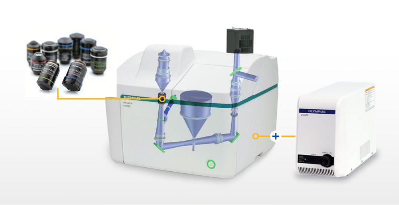
Advanced Optical Technology
Our advanced optical technology gives you the flexibility to capture high-quality images in many research applications.
- Uses leading-edge Olympus optics—including our award-winning X Line and silicone immersion objectives
- Six-position revolving nosepiece enables observation at multiple magnifications with the click of a button
- Uses a high color rendering LED for outstanding transmitted light imaging and a broadband LED light source for fluorescence
- Add a high-quality monochrome camera, and see fine details with accurate color rendering
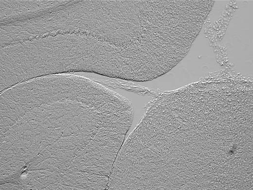
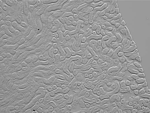
Gradient Contrast
Acquiring sharp images at the edges of a well in a multi-well plate can be challenging. The contrast is poor even when using oblique contrast, and DIC does not work well with plastic containers. Using our unique gradient contrast method, you can capture sharp images in a wider area and with higher contrast than the conventional method.
- Works with any Olympus objective
- Less affected by the meniscus, container lids, and water droplets
- Can be used with glass- and plastic-bottom dishes and multi-well plates
- Image through the plastic lids of Petri dishes and multi-well plates, reducing the risk of contamination
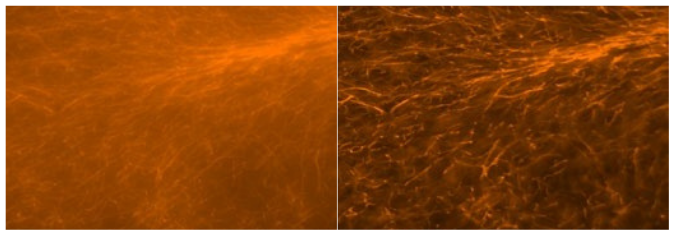
Clearer Images with Motorized Spherical Aberration Correction
Account for the thickness of your cover glass quickly and simply with the system’s motorized spherical aberration correction.
- No more manual correction collar adjustments
- Motorized adjuster is easy to set up and control
- Choose presets for glass or plastic containers or make custom adjustments
Images Are Organized and Easy to Find
The APX100 microscope has a dedicated system to organize and store your data. When you acquire an image, the software automatically creates folders for each sample and saves the data into the correct one. The consistent indexing keeps your data organized and easy to locate.
Recall Image Acquisition Settings
The APX100 system saves all important acquisition settings with the image data, making it easy to recall experiment conditions.
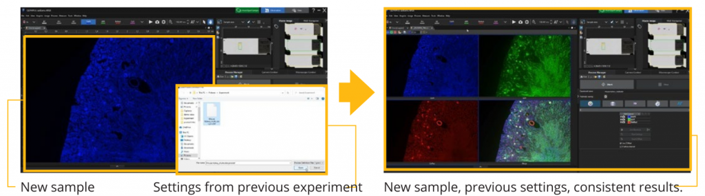
Image Processing and Analysis Functions that Accelerate Research
Our advanced cellSens image processing and analysis software works seamlessly with the APX100 system, providing you the tools to process and analyze your data quickly and efficiently.
cellSens Image Analysis
An optional cellSens software license provides powerful analysis capabilities.
- Access image processing techniques, including counting intracellular signals, analyzing the brightness of time-lapse data, and Olympus’ TruAI deep-learning technology
- The TruAI deep-learning solution offers flexibility, from predicting multi-class nuclei phenotypes for drug testing to rapid automated detection and segmentation of glomeruli
- cellSens integration enables easy, on-the-spot processing for simple measurements and image export
| Microscope | Observation method | Brightfield, fluorescence, phase contrast, gradient contrast |
| Sample holder | Glass slides (3 slides), 35 mm dish (3 dishes), microplate, general | |
| Objectives | Choose from 25 available objectives (4X–100X)
|
|
| Motorized aberration correction | One motorized position, five standard positions for objectives | |
| Transmitted illuminator | Built-in Köhler illumination for transmitted light, high color rendering LED
|
|
| Stage | Motorized XY stage with automatic control | |
| Focusing | Motorized focusing with automatic control | |
| Magnification changer | Color/Monochrome 0.5x fixed | |
| Fluorescence | Fluorescence illuminator with fly-eye lens
|
|
| Macro optical system | Built-in, 0.07x macro optics | |
| Anti-vibration mechanism | Built-in | |
| Camera | Color camera | 6.41 megapixels, 1/1.8 color CMOS |
| Monochrome camera | 6.41 megapixels, 1/1.8 monochrome CMOS | |
| Optional units | Stage-top incubator | Tokai Hit STX series (APX dedicated model) |
| Solution administration system | Tokai Hit KSX-Type2 | |
| Environment | Weight | 34.6 kg (76.3 lb) |
| Power consumption | 70 W
|
|
| Power supply ratings | Input : AC 90–264 V, 50–60 Hz
|
For other products from Evident-Olympus, click here.