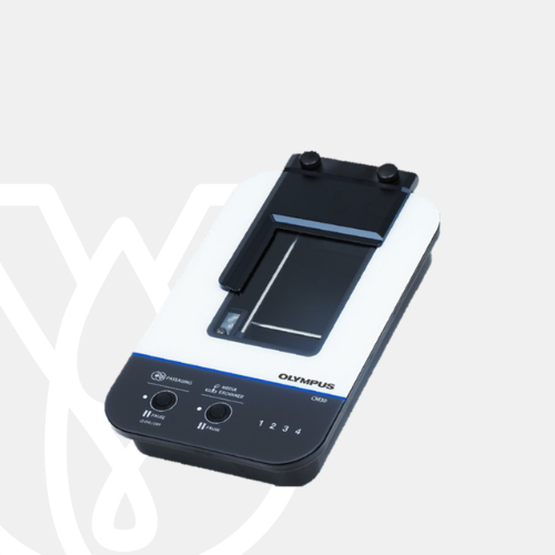
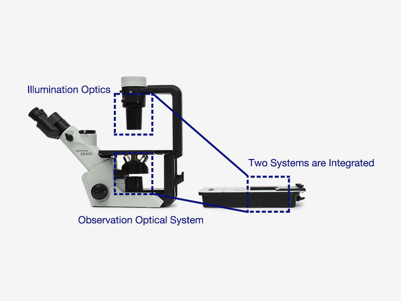
Olympus Incubation Monitoring System CM30
PT Wadya Prima Mulia sebagai Distributor Tunggal dari Evident-Olympus di Indonesia, menyediakan Sistem Monitoring Inkubasi CM30 dan produk lainnya.
Kontrol Proses Anda dengan Monitoring Kultur Sel Cerdas
Proses kultur sel merupakan proses yang mahal, rumit, dan memakan waktu. Dengan sistem monitoring inkubasi CM30, proses kultur dapat ditingkatkan dengan cara yang mudah.
Tingkatkan Prose Kultur Sel— Tanpa Label, Hasil Kuantitatif
Monitoring Sel Tanpa Label
Dengan sistem CM30, pewarnaan kultur tidak diperlukan untuk memonitor kondisinya. Sistem memperoleh data kuantitatif dari sel tanpa label, mengurangi potensi kerusakan pada kultur Anda.
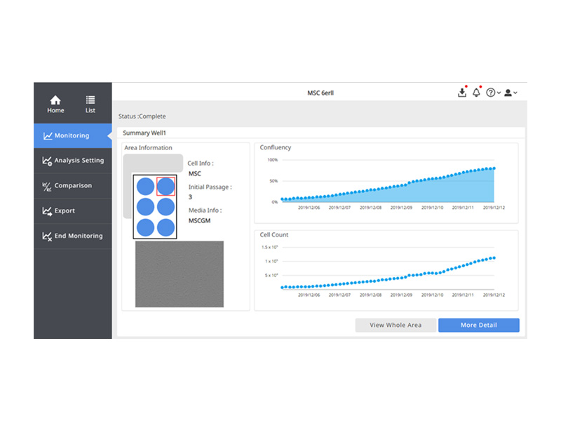
Monitoring Sel Kultur pada Berbagai Titik
Sistem secara otomatis memindai beberapa titik di wadah kultur Anda, memberikan data kuantitatif berkala tentang kesehatan dan konfluensi kultur Anda.
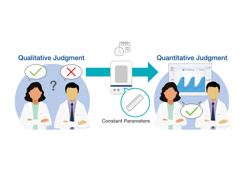
Hasil Konsisten Sepanjang Penelitian
Parameter Analisis Konstan
Sistem CM30 menggunakan teknologi analisis gambar berdasarkan machine learning untuk terus mengukur dan menganalisis gambar yang diperoleh. Memvisualisasikan status kultur sebagai nilai kuantitatif secara kontinu menghilangkan faktor yang menyebabkan variasi dalam pemeriksaan sel dan berkontribusi pada reproduktivitas dan konsistensi percobaan.
Membandingkan Data dari Berbagai Sampel
Menggunakan banyak head membantu Anda mempertahankan sampel kontrol sementara beberapa protokol pengujian berjalan pada waktu yang bersamaan. Bagikan data Anda dengan anggota tim atau bandingkan dengan data sebelumnya.
Sesuaikan Parameter Analisis agar Sesuai dengan Penelitian Anda
Sistem CM30 secara otomatis melakukan pertemuan, jumlah sel, dan jumlah koloni dari gambar yang diperoleh. Anda dapat mengonfigurasi parameter analisis sistem agar sesuai dengan kondisi setiap kultur sel, seperti jenis sel, kondisi kultur, atau pemberian obat. Mengetahui status kultur sel bertahap pada setiap waktu meningkatkan akurasi percobaan.
Efektif Biaya
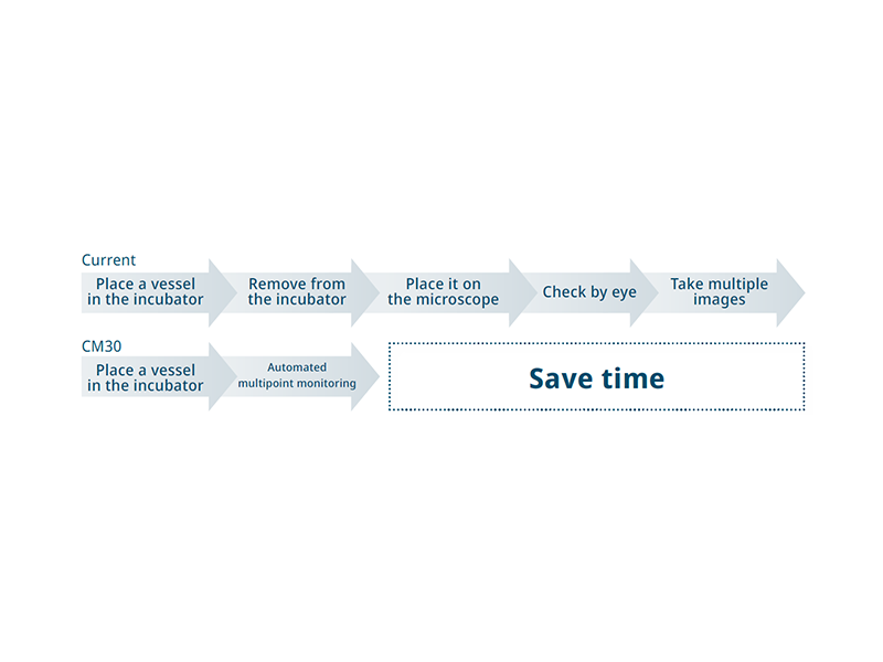
Menghemat Waktu dengan Automatisasi Sistem
Tingkatkan alur kerja berbasis mikroskop tradisional Anda dan dapatkan hasil yang lebih akurat dalam waktu yang lebih singkat. Dengan mengotomatiskan pemantauan kultur sel menggunakan sistem CM30, Anda dapat memperluas penelitian dan menggunakan waktu dengan lebih efektif
Tidak Perlu Masuk Ruangan Steril untuk Monitoring
Setiap memasuki ruang bersih, ada biaya operasional untuk bahan habis pakai dan pengukuran. Sekarang, Anda tidak perlu lagi selalu memeriksa status kultur sel di luar inkubator. Anda dapat mengurangi biaya dengan memeriksa status kultur Anda dari jarak jauh dari luar lab.
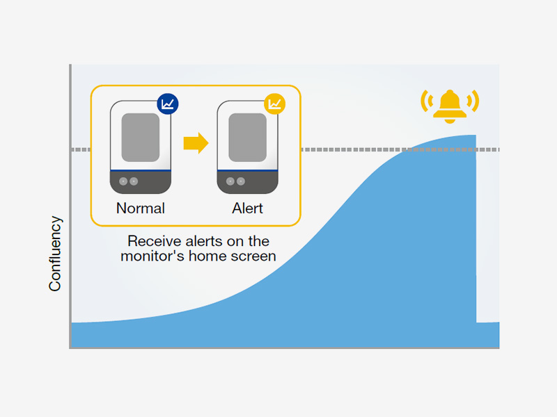
Menentukan Waktu Pasase Sel secara Akurat
Menentukan waktu pasase sel secara konsisten dan tanpa subjektivitas yang terkait dengan penghitungan manual. Berdasarkan parameter standar yang Anda tetapkan, perangkat lunak dapat menunjukkan kapan sel Anda siap untuk dilewati, membantu mencegah kegagalan.
Testimonials

Tokyo Medical and Dental University
Institute of Research
Banyak peneliti mungkin ingin tahu tentang mekanisme biologis yang terlibat dalam culturing jangka panjang yang diperlukan untuk pembentukan sel punca dan organoid di dalam inkubator. Sejujurnya saya terkejut bahwa mikroskop sederhana siap untuk menjawab pertanyaan ini karena CM20* ini dapat memvisualisasikan pembentukan spheroid dan organoid yang dibiakkan dalam gel matriks ekstraseluler dengan kontras yang menakjubkan. Saya percaya bahwa CM20 dapat menjadi alat transformatif dalam berbagai eksperimen biologi sel.

Translational Cardiovascular
Research Center University of Arizona
Dr. Erik Blackwood dan timnya telah menerapkan sistem pemantauan inkubasi CM20 untuk mengoptimalkan proses pemantauan kultur sel mereka. Mereka menggunakan sistem untuk menentukan kapan garis sel siap untuk uji praklinis pada model hewan pengerat untuk gagal jantung. Pendekatan mereka berfokus pada mengidentifikasi terapi berbasis obat molekul kecil, tetapi juga pada pendekatan terapi gen. Kemampuan penghitungan jumlah dan konfluensi dari sistem CM20 memungkinkan para peneliti memantau efek terapi ini pada garis sel iPS (induced pluripotent stem) secara real time sementara kultur tetap aman di dalam inkubator. Memanfaatkan fungsi pengumpulan data otomatis sistem juga meningkatkan efisiensi alur kerja mereka.
Pelajari bagaimana Dr. Blackwood dan timnya meningkatkan alur kerja kultur sel mereka dengan sistem monitoring CM20*.

Mr. Yoshihito Tachi
Saya bertanggung jawab atas penelitian dan pengembangan kit kondrosit autologus dan melakukan kultur sel dalam berbagai kondisi saat memeriksa protokol kultur. Saya sebelumnya menggunakan mikroskop untuk mengamati kondisi sel yang dikultur, tetapi saya hanya dapat melakukan evaluasi kualitatif dengan metode ini. Tantangan lainnya adalah hasil analisis bervariasi tergantung pada pengalaman dan subjektivitas pekerja. Sebaliknya, sistem Olympus CM20* dapat menghitung sel dan mengukur konfluensi dengan parameter analisis konstan yang sama untuk hasil analisis yang sangat dapat direproduksi dan konsisten. Kami dapat membandingkan data di antara eksperimen saat mengubah kondisi budaya, membandingkannya dengan data sebelumnya, dan membagikannya dengan mudah di dalam tim— yang semuanya dapat membantu kami meningkatkan efisiensi pengembangan. Kami juga mempertimbangkan untuk menggunakannya sebagai alat pendidikan. Dengan melihat indikator-indikator seperti tingkat perlekatan, keseragaman, dan kemampuan proliferatif, kita dapat mengevaluasi keterampilan pekerja dengan lebih baik.
*Review pengguna di atas dibuat setelah menggunakan CM20, tetapi hasil yang sama dapat diperoleh dengan menggunakan CM30.
Tidak Perlu Mengeluarkan Kultur dari Inkubator
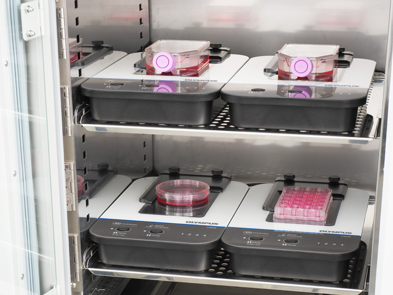
Monitor memungkinkan Anda melacak kesehatan kultur sel tanpa mengeluarkannya dari inkubator, mengurangi risiko kontaminasi atau kerusakan akibat perubahan suhu dan getaran. Desainnya yang unik memungkinkan Anda memasukkan hingga empat head unit di dalam inkubator standar untuk efisiensi yang lebih besar.

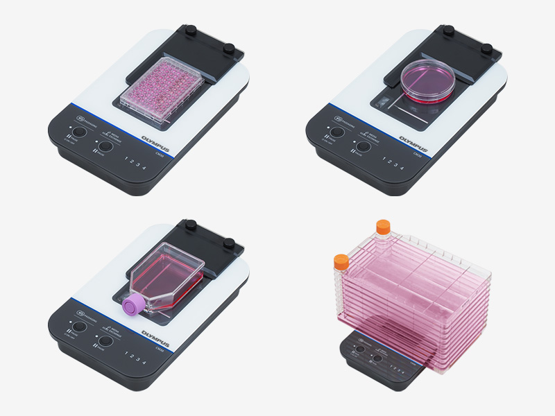
Desain Flat Mengakomodasi Berbagai Wadah
Sistem optik epi-oblique Evident memungkinkan sistem pemantauan inkubasi CM30 memiliki desain datar dan ringkas yang mengakomodasi sebagian besar bejana kultur sel standar. Cukup tempatkan bejana kultur yang biasa Anda gunakan pada CM30.
Analisis Sel Otomatis Menggunakan AI
Dengan melatih sistem untuk membedakan antara sel dan latar belakang menggunakan machine learning, secara otomatis dapat menentukan konfluensi.


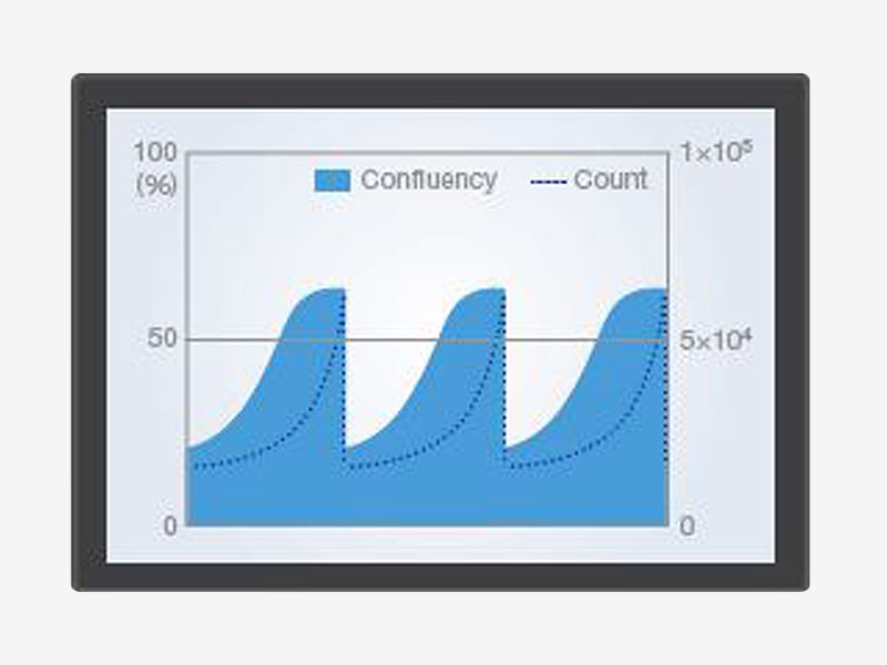
Data yang dapat Direproduksi dan Dibandingkan
CM30 memudahkan untuk menyimpan dan berbagi catatan kuantitatif terperinci tentang pertumbuhan dan kesehatan sel.
- Membuat grafik data konfluensi untuk melihat jumlah sel dan analisis koloni dalam urutan kronologis untuk melihat bagaimana perubahannya dari waktu ke waktu
- Fungsi perbandingan memudahkan untuk membandingkan data saat ini dengan hasil sebelumnya atau untuk membandingkan antar sumur
- Memungkinkan Anda dengan cepat menghitung data karakterisasi sel, seperti waktu penggandaan dan tingkat penggandaan populasi (PDL)
Sesuaikan Pengaturan untuk Memenuhi Kebutuhan Anda
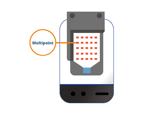
Titik Monitoring
Tetapkan titik pemantauan di wadah dan pelat sumur di mana saja yang Anda inginkan, seperti yang ditunjukkan di bejana di sebelah kanan. Dengan menentukan baris dan kolom, Anda dapat menetapkan beberapa titik sekaligus.
Posisi Fokus
Bebas mengatur posisi fokus untuk setiap titik pemantauan.
Waktu Monitoring
Bebas mengatur waktu mulai dan waktu interval.


Application Programming Interface (API) untuk Mengontrol Parameter Akuisisi Data (β ver.)
Kontrol head CM30 jarak jauh dan independen untuk:
Menentukan Parameter Kunci
- Pencahayaan
- Stage (XY, Z)
- Fokus (fokus auto/fokus manual)
- Eksposur dan pengambilan gambar
Pengambilan dan Akuisisi Gambar
- Pengambilan gambar statis
- Preview live
- Mengambil dan menghapus data gambar
- Mendapatkan daftar gambar
Layanan Validasi IQ/QQ
Tim dukungan kelas dunia kami dapat membantu Anda mengintegrasikan sistem pemantauan inkubasi CM30 ke dalam lab Anda. Layanan validasi IQ/OQ juga tersedia.

CM30H: Incubation Monitoring Head
| Installation environment (inside the incubator) | Temperature: 37 °C (98.6 °F) + 0.3 °C (0.5 °F), humidity: 0—99% |
| Applicable vessels | Petri dish (90 mm (3.54 in.), 100 mm (3.94 in.)) |
| Microplate (6 well, 12 well, 24 well, 48 well, 96 well) | |
| Flask (T25, T75, T80, T150, T175, T225) | |
| Multi-layer flask | |
| Optical performance | Field of view (H × V): 2.84 mm × 2.13 mm (0.11 in. × 0.08 in.); (image size per one shooting) |
| Image size: 1280 × 960 pixels | |
| Illumination wavelength: λ = 630 nm (LED) | |
| Illumination method: epi-oblique illumination | |
| Cable length | Approx. 4.5 m (14.8 ft) |
| Sterilization resistance | Autoclave sterilization (for vessel holder and sponge rubber only) |
| UV ray sterilization | |
| Hydrogen peroxide (H₂O₂) gas sterilization (CM30H only) | |
| Disinfection resistance | Peracetic acid disinfection (cold sterilant) |
| Alcohol disinfection | |
| Weight | Approx. 3.1 kg (6.8 lb) |
Incubation Monitoring Station
| Number of connectable CM30H | Max. 4 heads |
| HDD capacity | 4 TB or more |
Software
| User Management | 1000 user licenses (max) | |
| Project setting | Project creation: new or load | |
| Settings: standard or custom | ||
| Culture conditions: vessel information, culture information etc. | ||
| Cell analysis conditions: new or load | ||
| Access authority: public or private | ||
| Imaging interval: selection type | ||
| Analysis | Cell analysis: cell confluency, cell count | |
| iPS/ES cell analysis: colony confluency, colony count, colony size | ||
| Data statistics: growth rate, doubling time | ||
| Browsing | Image: entire area (tiling), fixed points | |
| Analysis result: graph (time, passage) | ||
| Export | Data export: image file, movie file* CSV file* *only for fixed points | |
| Create report (PDF) |
Client PC (Recommended System Configuration for CM30 Software)
| OS | Microsoft® Windows® 10 (64-bit) or higher |
| CPU | Intel® Core TM i3 (2.1 GHz) or more |
| RAM | 4 GB or more |
| HDD | Free space: 2 GB or more |
| Screen resolution | 1366 × 768 or more |
| Web browser | Google Chrome TM |
Operation Confirmed Incubator
| Thermo Fisher Scientific | 51030388 |
| Panasonic | MCO-170AICUVH-PJ |
| ASTEC | SMA-165DRS |
| Eppendorf | CellXpert® C170i (Operation confirmed by Eppendorf)
Reference data:Remote and quantitative cell culture monitoring: Vero cells |
Incubator conditions required for use of the CM30 system:
- The minimum height of the CM30H installation space must be 150 mm (6 in.)
- There must be an access port through which the CM30H cable can be taken out
Untuk produk lain dari Evident-Olympus, klik disini.