Olympus Research Slide Scanner VS200
PT Wadya Prima Mulia as the Exclusive Distributor for Evident-Olympus in Indonesia, provides VS200 Research Slide Scanner and other products.
Easily analyze, share, and archive your data with the SLIDEVIEW VS200 research slide scanner. Designed to capture high-resolution images of your slides for quantitative analysis, the system enables you to make the most of the information your slides have to offer.
Outstanding Image Quality for Quantification
Better Resolution and Flatness
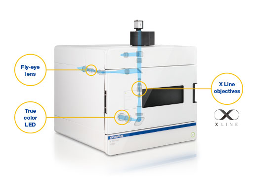
- Uses X Line high-performance objectives to deliver images with no stitching artifacts or patchwork effect.
- An optimized optical beam path provides more homogenous illumination.
- High-power True Color LED matches the spectral performance of reference halogen light sources, so brightfield stains like purple, cyan, and pink are correctly represented, imaged, and rendered.
- Fluorescence illuminator with a fly-eye lens uniformly distributes light for bright, even images.
- Optional optical sectioning device produces high-contrast images, especially for thick samples.
Flexible for Many Applications
Supports Multiple Hardware Options
The VS200 slide scanner is a highly flexible system that supports various slide sizes, different objectives, the SILA optical sectioning device, oil dispenser, cameras, loaders, and LED/laser light sources with multiple filter sets.
Supports Multiple Observation Methods
The VS200 supports brightfield, darkfield, polarization, phase contrast, and fluorescence, with the possibility to scan a sample using multiple techniques at the same time.
Flexible Software Solutions
The VS200 software supports offline analysis, deep learning, 3D deconvolution, and batch conversion to standard file formats. It also supports the NIS-SQL database for data management and the DICOM format for easy integration into a lab information management system (LIMS).
Achieve More in Less Time
- Loader holds up to 35 sample trays with a maximum capacity of 210 26 × 76 mm (1 × 3 in.) slides
- Robotics quickly and safely loads and unloads trays while coordinating with the focus and scan unit for fast acquisition
- Integrated barcode reader automatically captures and records slide information
Simplified and Powerful Workflow
- Simple user interface—choose expert mode to customize the system’s settings or quick mode where the software optimizes the settings for you
- Save and recall settings to speed up repetitive workflows and standardize operations
- Settings can be saved as projects and shared among multiple users
- TruAI technology—post-processing deep learning enables accurate segmentation of morphological features on a sample
Reliable Data for Many Applications
The flexibility of five imaging modes and outstanding Olympus X Line objectives make the VS200 slide scanner a solution for many applications.
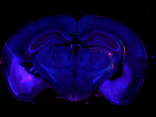
Brain Research
Cortico-thalamic projection pathways labeled with AAV-GFP and AAVtdTomato.
Image data courtesy of Hong Wei Dong, MD, PhD, Professor of Neurology, Keck School of Medicine of University of Southern California.
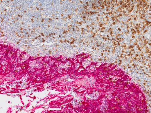
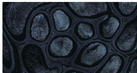
Cancer and Stem Cell Research
Tonsil CD3 (rm), ImmPRESS Reagent (HRP) Anti-Mouse IgG Immpact DAB (brown), AE1/AE3(m) ImmPRESS (AP) (HRP) Anti-Rabbit IgG Immpact Vector Red (red). Counterstained with Hematoxylin QS (blue).
Image data courtesy of Vector Labs.
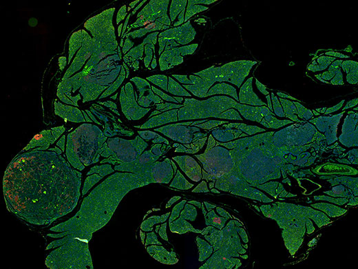
Drug Discovery
Pancreas stained with Dapi, GFP and RFP.
Image data courtesy of Wenjin Chen, NJ Rutgers Cancer Center.
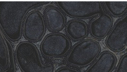
Accurate Focus at All Magnifications
- Z-focus is 4X more precise than our previous slide scanner and offers higher readjustment accuracy with linear encoders
- Automatic sample detection helps determine the scan area for faster imaging while the autofocus improves the automated stitching function
- Design minimizes vibrations for better imaging stability
- Manually edit the sample mask and modify the automatically detected sample mask
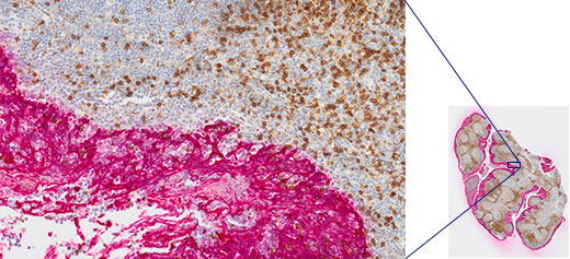
Image data courtesy of Vector Labs.
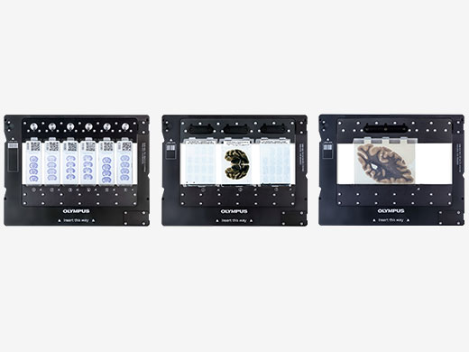
Supports Glass Slides and Plates
- Simple-to-use slide tray enables you to mix 26 × 76 mm (1 × 3 in.), 52 × 76 mm (2 × 3 in.), 76 × 102 mm (3 × 4 in.), and 102 × 127 mm (4 × 5 in.) slides on the same batch scan
- A RFID system enables the unit to automatically detect trays, the number of slides, and the size of the slides
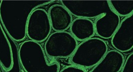
The Power of TruAI Deep-Learning Technology
- Deep learning enables faint samples or morphological features on a sample to be accurately identified and scanned, saving time and effort
- Target regions, such as pancreas islets, can be accurately segmented and differentiated from erythrocytes
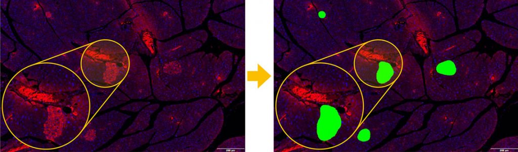
Right: Probability map detection based on TruAI technology. Only pancreatic islets are accurately detected (green).
Image data courtesy of Univ.-Prof. Dr. rer. nat. Simone E. Baltrusch, Institute for Medical Biochemistry and Molecular Biology, Rostock University Medical Center, University of Rostock.
| Intended Specimen | Observable Specimen | Glass slide with cover glass |
| Size of Glass Slide (W × L × H) |
Standard slide tray: 25 mm–26.5 mm × 75 mm–76.5 mm × 0.9 mm–1.2 mm (1 in. × 3 in. × 0.05 in.) (6 slides)
| |
| Cover Glass Thickness | 0.12–0.17 mm | |
| Observation Methods | Brightfield, reflected brightfield (optional*1), darkfield, phase contrast (optional*2), simple polarization (optional*3), fluorescence (optional), fluorescence optical sectioning with speckle illumination (optional SILA module) | |
| Illuminator | Zoom Ratio | Built-in Köhler illumination for transmitted light; high intensity and high color rendering LED (up to 50,000 hours) |
| Objectives | Compatible objectives 2x, 4x*4, 10x*5, 20x, 40x*5, 60x*5, and 100x*5 6-position motorized nosepiece (incl. selected oil immersion, silicon oil immersion, and phase contrast objectives) Optional automatic oil dispenser | |
| Motorized Stage | XY stage with automatic control | |
| Focusing | Motorized focusing with automatic control | |
| Color Camera | Integrated 2/3 inch CMOS, 3.45 μm × 3.45 μm pixel size, high sensitivity, high resolution | |
| Scan Unit | Capacity | 1 slide tray, 6 slides maximum; upgradable to a multiple tray loader model |
| Pixel Resolution(Color Camera) |
UPLXAPO20X (NA 0.8): 0.274 μm/pixel
|
|
| Scan Time | Brightfield: approx. 1.5 minutes (20x objective, scan area 15 mm × 15 mm) Fluorescence widefield NOVEM: approx. 6.5 minutes (20x objective, scan area 15 mm × 15 mm, 4 fluorescence channels, 10 ms exposure each) | |
| Software | Automatic sample detection (generic and TruAI deep learning), automatic barcode reading, automatic focus mapping, automatic scanning, automatic stitching, pause and resume scanning, Z-stack imaging, extended focus imaging (EFI), image format: vsi, JPEG, TIFF, DICOM, synchronized multi-image display, stepless zooming, zooming while scanning, annotations, screen capture, slide loader control (multiple tray loader only) | |
| Fluorescence (optional) | Fluorescence Components |
UPLFLN4X objective, illuminator with fly-eye lens, motorized mirror turret, motorized filter wheel
|
| Monochrome Camera |
Options:
|
|
| Solutions for Scanner Software (optional) | Solution License |
Batch image format converter
|
| Desktop Software (optional, separate solution for analysis) | Solution License |
Batch image format converter
|
| Environment | Weight | Optical frame: 69 kg (152.1 lb)
|
Fluorescence: 8 kg (17.6 lb)
|
| Operating Environment | Temperature: 15–28 °C (59–82.4 °F) (including other devices)
|
|
| Power Supply | Input: AC 100–240 V, 50/60 Hz, 4 A
|
|
| Power Consumption | Input: 100–240 V AC; 50/60 Hz; 4 A 221 W |

For other products from Evident-Olympus, click here.