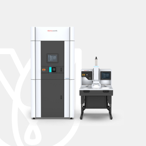
PT Wadya Prima Mulia as the Authorized Distributor for ThermoFisher Scientific in Indonesia, provides Tundra Cryo-Transmission Electron Microscope
The Thermo Scientific Tundra Cryo-Transmission Electron Microscope (Cryo-TEM) allows you to perform structural analyses on challenging proteins and macromolecules with unprecedented ease of use, at a price point within the reach of many grants and funding sources.
For more information regarding the product, click here
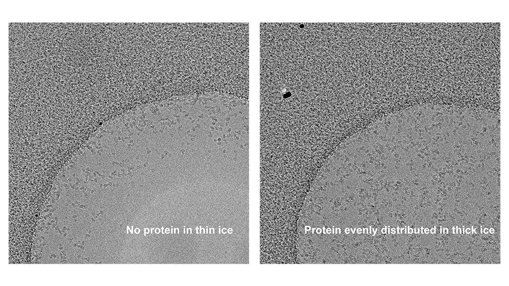
Shorten time to cryo-EM sample selection
The Tundra Cryo-TEM can visualize the impact of biochemical adjustments to samples faster than other technologies. Exchange samples in and out of the microscope in minutes, with nearly instantaneous feedback, allowing you to optimize your sample conditions quickly without extensive training.
Structural information at biologically relevant resolution
The optics of the Tundra Cryo-TEM are specifically designed so that you can answer fundamental questions on biology and human disease with data at biologically relevant resolutions. Operating at 100 kV, the Tundra Cryo-TEM can generate high-contrast biological images routinely at near atomic resolution with medium throughput with the high performance Thermo Scientific Falcon C Direct Electron Detector.
Designed to meet your budget
A wide range of funding proposals and grants were considered when designing the Tundra Cryo-TEM to keep its price within the reach of most instrumentation grants. Our Electron Microscopy Funding Support Center has additional information about our financial services as well as grant-writing guidance so you can get the tools you need, when you need them.
Advanced cryo-TEM software takes the guesswork out of cryo-TEM
The Tundra Cryo-TEM comes with a complete suite of automation software for efficient optimization of your sample’s biochemistry as well as data collection for structural determination. This includes a user-friendly AI-enabled software solution capable of analyzing intermediate results, providing instant feedback, and steering data collection on the fly. Our AI algorithms are based on years of cryo-EM knowledge, providing expert decisions upfront to ensure that your instrument is working at optimal conditions. This allows you to focus on the science rather than on fine-tuning the microscope.
Tundra Cryo-TEM features
The Tundra Cryo-TEM is equipped with a 100 kV high-brightness field emission gun (X-FEG) that allows for high resolution structure determination at routinely 3-5 Å. The Tundra Cryo-TEM can be installed with either the Falcon C Direct Electron Detector or Thermo Scientific Ceta F CMOS Detector. The Falcon C Direct Electron Detector provides a boost in contrast for single particle cryo-EM, improving resolution and obtaining results faster with less data to maximize your resolution efforts. Tundra Cryo-TEM with the Falcon C Direct Electron Detector makes it possible to efficiently solve protein structures 100 kDa or less.
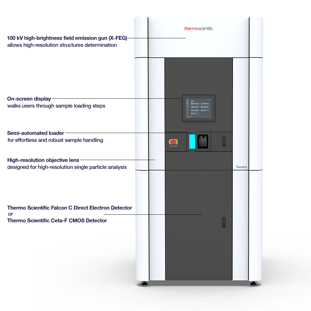
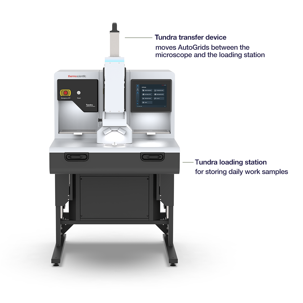
Tundra Cryo-TEM loading station
An integrated loader makes it easier for users of any experience level to load samples into the microscope, as compared to conventional systems. Scientists can exchange sample carriers in minutes. Results can be checked immediately, allowing users to rapidly optimize biochemistry sample conditions.
The cryo-loading station stays cold for approximately 6.5 hours. With a 20-minute loading and unloading cycle, as well as time for sample quality analysis, users can exchange up to 10 sample grids a day.
| Source | X-FEG (extreme high-brightness field emission gun) |
| Accelerating voltage | Fixed accelerating voltage of 100 kV |
| Semi-automated sample loading | • Cryo-preparation station for contamination free sample exchange • Sample transfer device for transfer of single AutoGrids to the microscope, with fixed cryo-box |
| Lenses | High-resolution objective lens optimized for single particle analysis |
| Stage | Computerized specimen cryo-stage |
| Imaging options | • High-performance Falcon C Direct Electron Detector for improved resolution capabilities • Standard Ceta-F CMOS Camera with dose fractionation, optimized for low-dose imaging |
| Additional components | Primary control unit with two 24” monitors to be placed within 10 m of the column |
| Software | AI-enabled and automated software solution that provides feedback during data collection and has an open application programming interface |
With the Thermo Scientific Tundra Cryo-TEM, configured with the Thermo Scientific Falcon C Direct Electron Detector, you can visually resolve protein structures and produce 3D reconstructions down to 2.1 Å. The Tundra Cryo TEM can also be utilized for negative-stain electron microscopy, an easy and cost-effective method for the quality assessment of purified biological specimens at room temperature. In addition, the Tundra Cryo-TEM can visualize sections of resin-embedded cells and tissues or isolated particles of protein complexes and viral assemblies.
Single particle cryo-EM analysis
The 100 keV Tundra Cryo-TEM can efficiently validate ligand and protein binding when in fast mode, taking over 10,000 images in as little as a few days’ acquisition time. With the Tundra Cryo-TEM, you can take a closer look at receptors that affect the central nervous system or investigate prokaryotic ribosomes. From Nobel –prize-winning discoveries, like TRPV5, to small potential drug targets, like GABAA receptors, the Tundra Cryo-TEM can help expand your scientific discovery by visually resolving structures and producing 3D reconstructions at ~2–5 Å resolution. When the Tundra Cryo-TEM is configured with a Falcon C Direct Electron Detector, the resolution can be improved, particularly for small and asymmetric protein complexes such as transthyretin (55 kDa).
Drug discovery with the Tundra Cryo-TEM
Cryo-electron microscopy (cryo-EM) is rapidly drawing interest in structure-based drug discovery and design, since it can accurately and rapidly visualize to high resolutions the interactions between drug and receptor of a multitude of samples. Here we show an example of a human CDK-activating kinase (CAK), a challenging protein for traditional structural biology methods. Structures determined on the Tundra Cryo-TEM provide insight into inhibitor interactions and the basis for rational design of next-generation therapeutics.
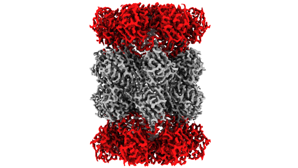
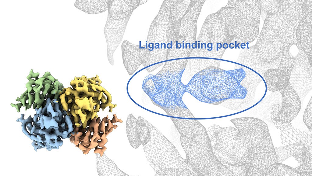
Reconstruction map of human CDK-activating kinase
The reconstruction map of the human CDK-activating kinase (CAK) was obtained at 4.3 Å resolution using the Tundra Cryo-TEM and Falcon C Direct Electron Detector. The structural insights at this resolution allowed researchers to determine how large ligands bind to the protein: a promising target for cancer therapeutics. The Tundra Cryo-TEM generated structure shows distinct CAK subunits: Cyclin H brown, MAT1 orange, CDK7 grey, AMP-PNP (an inhibitor of CAK) purple.

Cryo-TEM sample optimization and connectivity
The Tundra Cryo-TEM can be used to optimize samples, offering efficient sample iteration and optimization. It serves as a valuable screening tool, providing biochemical sample optimization in less time than higher-level cryo-EM instruments. In the example of CAK complex, when using Krios G4 Cryo-TEM, higher resolution reconstruction can be achieved to reveal more structural information.

Room temperature TEM applications
The Tundra Cryo-TEM can be utilized for negative-stain electron microscopy, an easy and cost-effective method for the quality assessment of purified biological specimens at room temperature to quickly assess the stability, homogeneity, and concentration of purified samples. In addition, room temperature sections of resin-embedded cells and tissues or isolated particles of protein complexes and viral assemblies can also be visualized with the Tundra Cryo-TEM.

Sample courtesy of New York Structural Biology Institute.

Sample courtesy of Sarah Powers, Doug Allen Lab, Janithri Wickramanayake, and Kirk Czymmek, Advanced BioImaging Laboratory, Donald Danforth Plant Science Center.
For other products from ThermoFisher, click here.