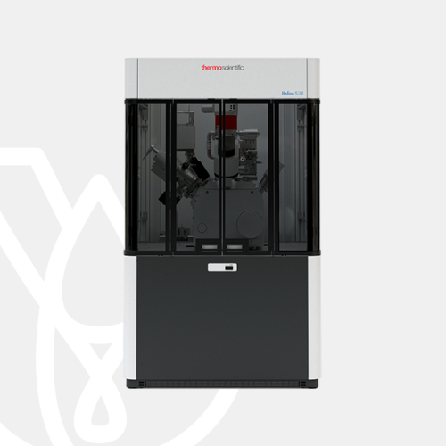
PT Wadya Prima Mulia as the Authorized Distributor for ThermoFisher Scientific in Indonesia, provides Helios 5 DualBeam
Sample preparation for TEM and STEM imaging or atom probe tomography. Easy to use with advanced automation. Capable of high quality subsurface 3D characterization.
For more information regarding the product, click here
FIB sample preparation
The new Thermo Scientific Helios 5 DualBeam builds on the high-performance imaging and analysis capabilities of the industry-leading Helios DualBeam family. It is carefully designed to meet the needs of materials science researchers and engineers for a wide range of focused ion beam scanning electron microscopy (FIB-SEM) use cases – even on the most challenging samples.
The Helios 5 DualBeam redefines the standard in high-resolution imaging with high materials contrast; fast, easy, and precise high-quality sample preparation for (S)TEM imaging and atom probe tomography (APT) as well as the high-quality subsurface and 3D characterization. Building on the proven capabilities of the Helios DualBeam family, additional advancements to the new Helios 5 DualBeam were designed to ensure the system is optimized for a variety of manual or automated workflows. Those improvements include:
- Greater ease-of-use: The Helios 5 DualBeam is the most accessible DualBeam for users of all experience levels. Operator training may be reduced from months to days and the system design is helping all operators to achieve consistent, repeatable results on a wide variety of advanced applications.
- Increased productivity: Advanced automation capabilities, increased robustness and stability enhancements in the Helios 5 DualBeam and Thermo Scientific AutoTEM 5 software can significantly increase the sample preparation throughput by allowing unattended and even overnight operation.
- Improved time to results: The Helios 5 DualBeam now includes FLASH, a new concept of tuning the image. With conventional microscopes, each time an operator needs to acquire an image, the microscope has to be carefully tuned by iterative alignments. With the Helios 5 DualBeam, a simple gesture across the screen will activate FLASH, which automatically adjusts these parameters. The automatic adjustments can significantly improve throughput, data quality, and simplify the acquisition of high-quality images.
TEM sample preparation
The Thermo Scientific Helios 5 DualBeam is part of the fifth generation of the industry-leading Helios DualBeam family. It is carefully designed to meet the needs of scientists and engineers, combining the innovative Elstar electron column for extreme high-resolution imaging and the high materials contrast with the superior Thermo Scientific Tomahawk Ion Column for the fast, easy, and precise high-quality sample preparation. In addition to the advanced electron and ion optics, the Helios 5 DualBeam incorporates a suite of state of-the-art technologies that enables simple and consistent high-resolution (S)TEM and atom probe tomography (APT) sample preparation, as well as the high-quality subsurface and 3D characterization, even on the most challenging samples.
High-quality sample preparation
Site-specific sample preparation for (S)TEM and APT analysis using the high-throughput Thermo Scientific Tomahawk Ion Column or the Thermo Scientific Phoenix Ion Column with unmatched low-voltage performance.
Fully automated
Fast and easy, fully automated, unattended, multi-site in situ and ex situ TEM sample preparation and cross-sectioning using optional AutoTEM 5 Software.
Shortest time to nanoscale information
For users with any experience level using best-in-class Thermo Scientific Elstar Electron Column featuring Thermo Scientific SmartAlign and FLASH technologies.
Next-generation UC+ monochromator technology
Reveal the finest details with the next-generation UC+ monochromator technology with higher current, enabling sub-nanometer performance at low energies.
Complete sample information
Sharp, refined and charge-free contrast obtained from up to six integrated in-column and below-the-lens detectors.
3D analysis
The high-quality, multi-modal subsurface and 3D information with the precise region-of-interest targeting using optional Thermo Scientific Auto Slice & View 4 (AS&V4) Software.
Rapid nanoprototyping
Fast, accurate and precise milling and deposition of complex structures with critical dimensions of less than 10 nm.
Precise sample navigation
Tailored to individual application needs thanks to the high stability and accuracy of the 150-mm piezo stage or the flexibility of the 110-mm stage, as well as the in-chamber Thermo Scientific Nav-Cam Camera.
Artifact-free imaging
Based on integrated sample cleanliness management and dedicated imaging modes such as DCFI and SmartScan Modes.
STEM imaging
The Thermo Scientific Helios 5 FX configuration offers a high productivity workflow with its unique, in-situ, 3Å resolution STEM capability.
Helios 5 CX
Workhorse preparation and XHR SEM imaging
| SEM resolution | 20 eV – 30 keV |
| SEM landing energy | • 0.6 nm @ 15 keV • 1.0 nm @ 1 keV |
| STEM Resolution @ 30 keV | 0.7 nm |
| FIB preparation process Max material removal | 100 nA |
| FIB preparation process Optimal final polish | 2 kV |
| TEM sample preparation Sample thickness | 50 nm |
| TEM sample preparation Automation | No |
| Sample handling Travel | 110 x 110 x 65 mm |
| Sample handling Loadlock | Manual Quickloader |
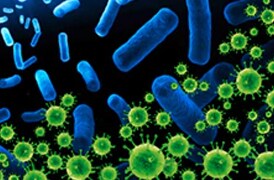
Cryo-EM techniques enable multiscale observations of 3D biological structures in their near-native states, informing faster, more efficient development of therapeutics.
Cryo-electron microscopy enables the structural analysis of challenging biological targets such as large complexes, flexible species and membrane protein.

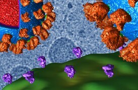
Learn how to take advantage of rational drug design for many major drug target classes, leading to best-in-class drugs.
Fundamental plant biology research is enabled by cryo electron microscopy, which provides information on proteins (with single particle analysis),
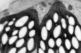
Transmission electron microscopy (TEM) is used when the nature of the disease cannot be established via alternative methods.
Process control using electron microscopy
Modern industry demands high throughput with superior quality, a balance that is maintained through robust process control. SEM and TEM tools with dedicated automation software provide rapid, multi-scale information for process monitoring and improvement.
Quality control and failure analysis
Quality control and assurance are essential in modern industry. We offer a range of EM and spectroscopy tools for multi-scale and multi-modal analysis of defects, allowing you to make reliable and informed decisions for process control and improvement.
Fundamental Materials Research
Novel materials are investigated at increasingly smaller scales for maximum control of their physical and chemical properties. Electron microscopy provides researchers with key insight into a wide variety of material characteristics at the micro- to nano-scale.
For other products from ThermoFisher, click here.