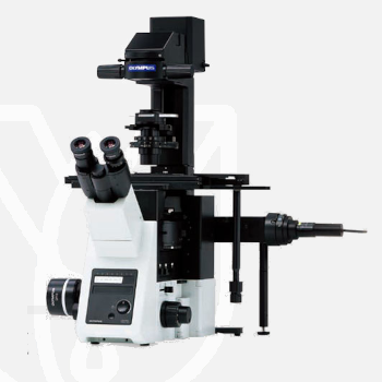
Olympus Inverted Microscope IXplore Standard
PT Wadya Prima Mulia as the Exclusive Distributor for Evident-Olympus in Indonesia, provides Inverted Microscope IXplore Standard.
High-Quality Imaging
Designed for simple multicolor fluorescence imaging and routine experiments, the IXplore Standard microscope system is easy to operate and capable of excellent, publication-quality images. A range of encoded unit options make it easy to get accurate and reproducible results.
- Repeatability and accuracy for standard imaging tasks
- Benefit from the same optical capabilities found in high-end IXplore systems
- Easily upgrade to encoded functionality to boost reproducibility of experiments
- Obtain high-quality images, even with standard cell culture vessels, using dedicated objectives
For more information about this product click here.
Accuracy and Repeatability
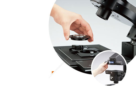
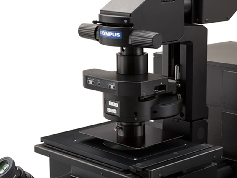
Smooth Tracking at High Magnification
The IX3-SVR manual stage features a smooth positioning system that enables cells to be easily tracked, even at high magnifications.
- Observation limit settings immobilize the stage and help maintain its position, including during reagent applications, even if the stage is inadvertently touched.
- Using the stage’s fixing plate and positioning screws, you can remove a 35 mm dish from the stage, transfer it to an incubator, and return it to its exact previous position.
Easy Köehler Illumination
The condenser can be moved and easily reset to Köhler illumination using the conveniently located condenser lock and control knobs.
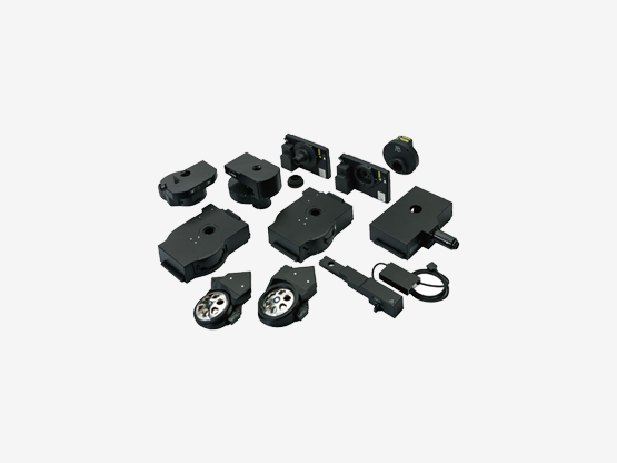
Encoded Units (Optional Peripherals)
A Cost-Effective Way to Upgrade to a Smart Microscope
A wide range of optional units are available for upgrades, including:
- 8-position motorized or encoded fluorescence mirror turrets
- 6-position motorized or encoded nosepiece
- Motorized long working distance universal condenser
- Filter wheels and shutters
Ease of Use
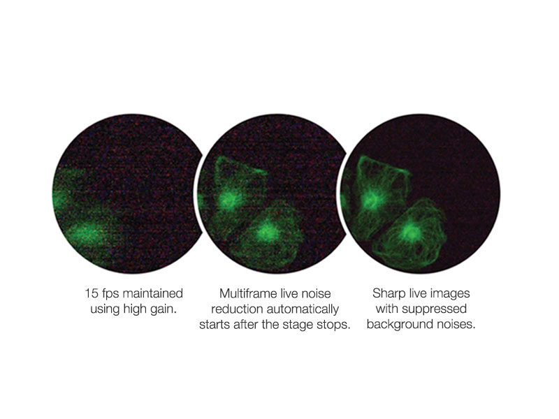
Focus on Dim Fluorescence Signals with Intelligently Designed Noise Reduction
The multiframe live noise reduction (FNR) feature makes it possible to capture low-noise, high-speed images of live specimens in real time. Olympus’ noise reduction technology enables users to obtain sharp fluorescence images, even in when fluorescence signal is low. The FNR feature takes multiple pictures of an image and then eliminates random background noise. The camera will stop the FNR automatically when the stage is moving, making it easier to scan your specimen and focus during fluorescence imaging.
High Contrast under Bright Conditions
The unit is designed specifically for fluorescence observation. It efficiently blocks out room light, enhances the contrast of fluorescence, and enables clear fluorescence observation under bright conditions.
Intuitive operation with cellSens imaging software
Olympus cellSens software is easy to use, powerful, and flexible. Using a modular design, the software can be tailored to your budget and imaging applications. This enables the software to grow and adapt to meet evolving research needs.
Simplify Your Workflow
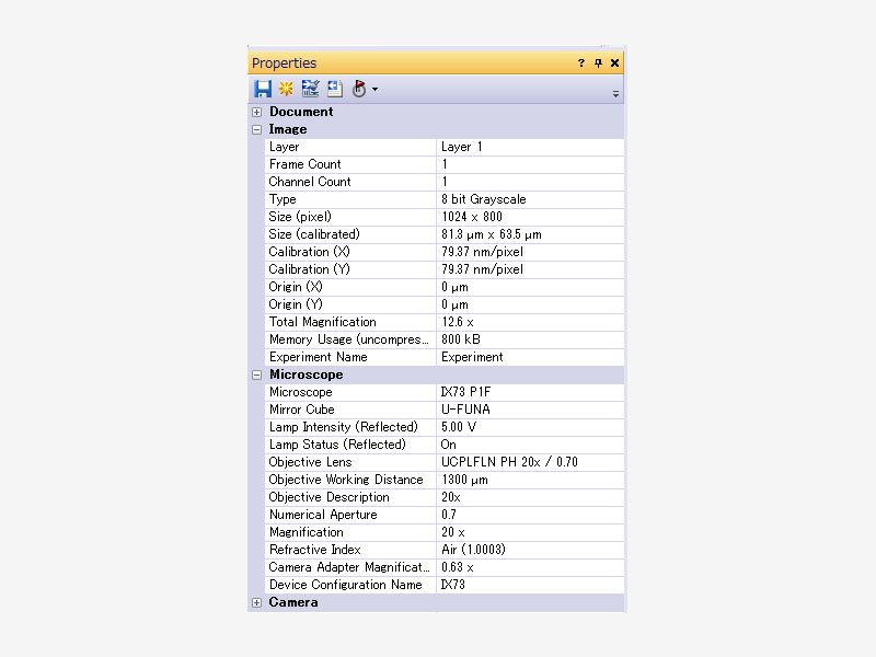
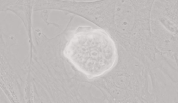
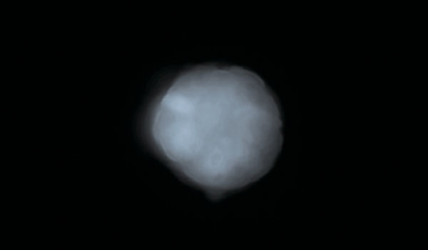
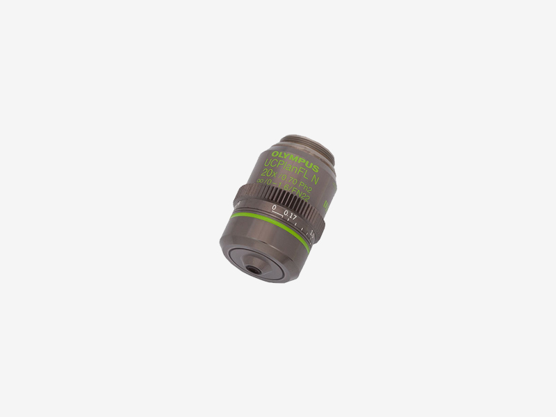
Objectives for Observation Using Plastic Vessels
LUCPLFLN series objectives, and in particular the UCPLFLN20XPH (NA 0.7), are well-suited for observation using plastic dishes. The objectives enable high-resolution observation of the cell proliferation process and deliver improved contrast across a wide area. This gives you the flexibility to image through plastic-bottom dishes in addition to glass.*Image: iPS-cell expressing Nanog reporter (GFP) Image data courtesy of: Tomonobu Watanabe, Ph.D. Laboratory for Comprehensive Bioimaging, RIKEN Quantitative Biology Center
High-Quality Imaging
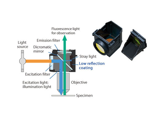
High Signal-to-Noise Fluorescence Mirror Units for Efficient Signal Detection
All fluorescence mirror units feature filters treated with a specially developed coating that absorbs more than 99% of stray light. This reduction in reflections and high transmittance of the mirror units helps provide fluorescence images with a high signal-to-noise ratio.
| Microscope Frame |
IX73P2F |
||
|---|---|---|---|
| Observation Method | Fluorescence (Blue/Green Excitation) | ||
| Fluorescence (Ultraviolet Excitation) | |||
| Differential Interference Contrast (DIC) | |||
| Phase Contrast | |||
| Brightfield | |||
| Revolving Nosepiece | Motorized (6 position) | ||
|
Revolving Nosepiece
|
Manual | Coded (6 position) | |
| Observation Tubes | Widefield (FN 22) | Tilting Binocular | |
| Illuminator | Transmitted Köhler Illuminator | LED Lamp | |
| 100 W Halogen Lamp | |||
| Fluorescence Illuminator | 100 W Mercury Lamp | ||
| Light Guide Illumination | |||
| Fluorescence Mirror Turret | Motorized (8 position) | ||
| Manual | Coded (8 position) | ||
| Stage | Motorized | IX3-SSU Ultrasonic Stage for IX3 |
|
| Mechanical | IX3-SVR Mechanical Stage with Right Handle |
|
|
| IX3-SVL Mechanical Stage with Left Short Handle |
|
||
| Condenser | Motorized | Universal Condenser | W.D. 27 mm, NA 0.55, motorized aperture and polarizer |
| Manual | Universal Condenser | NA 0.55/ W.D. 27 mm | |
| Ultra-Long Working Distance Condenser | NA 0.3/ W.D. 73.3 mm | ||
| Confocal Scanner |
– |
||
| Super Resolution Processing |
– |
||
| Accessories |
– |
||
| Dimensions (W × D × H) | 323 (W) x 475 (D) x 721 (H) mm (IX73 microscope frame) | ||
| Weight | Approx. 47kg (IX73P2F) | ||
For other products from Evident-Olympus, click here.